
CIE iGCSE Co-Ordinated Science B9.3 Blood vessels Exam Style Questions Paper 4
Question
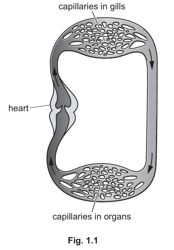
Explain the advantages of a double circulation compared to the circulation in a fish.
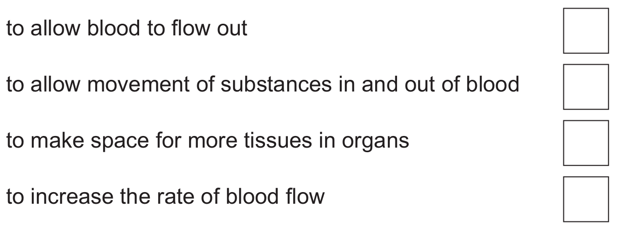
(i) Tick \((\checkmark)\) one box to identify which statement explains why capillaries have thin walls.
(ii) Fish gills have a good blood supply as they have many capillaries with thin walls. Suggest one other feature that makes the gills a good gas exchange surface.
An \(8\text{ mm}^3\) sample is taken from a patient.
The sample contains \(1.022\times 10^{6}\) platelets.
(i) Calculate the number of platelets per mm³ in the sample.
(ii) Suggest how this number of platelets may affect the patient if they cut themselves.
Most-appropriate topic codes (Cambridge IGCSE Co-ordinated Sciences 0654):
• Topic B9.1 — Circulatory systems (Parts (a) & (b))
• Topic B9.3 — Blood vessels (Part (c)(i))
• Topic B11.1 — Gas exchange in humans (Part (c)(ii))
• Topic B9.4 — Blood (Part (d)(i) & (d)(ii))
▶️ Answer/Explanation
(a) X should be placed on the right-hand side of the circulatory system (the region after the gills).
In fish, blood becomes oxygenated as it passes through the gills. The right side of the diagram shows oxygenated blood flowing from the gills to the organs.
(b) Three advantages of double circulation:
• Blood is circulated more quickly in the mammalian system.
• Higher blood pressure is maintained to supply the body tissues.
• Lower blood pressure is maintained for the lungs, preventing damage to delicate capillaries.
• More effective delivery of oxygen and nutrients to body cells.
• Oxygenated and deoxygenated blood are kept completely separate.
(c)(i) ✓ to allow movement of substances in and out of blood
Thin capillary walls (single cell layer thick) create a short diffusion distance, allowing efficient exchange of oxygen, carbon dioxide, nutrients, and waste products between blood and tissues.
(c)(ii) Large surface area / good ventilation with water.
Gills have many lamellae which increase surface area, and water flows continuously over them maintaining a steep concentration gradient for efficient gas exchange.
(d)(i) Number of platelets per mm³:
\(\frac{1.022 \times 10^6}{8} = 127750\) platelets per mm³
(Alternative: \(1.278 \times 10^5\) platelets per mm³)
(d)(ii) The platelet count is below the healthy range (127750 < 150000).
This may cause:
• Reduced blood clotting ability.
• Increased risk of pathogens entering the wound.
• Increased blood loss from the cut.
• Slower wound healing.
Question
Topic codes:
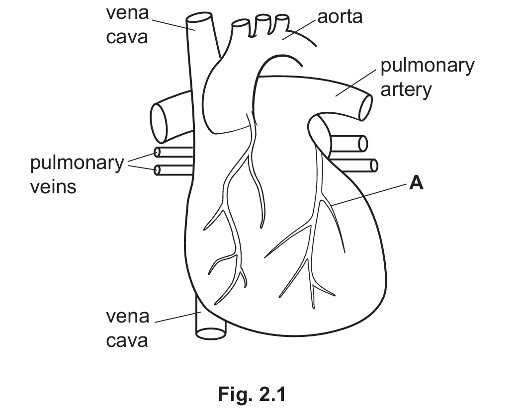
• Topic B9.2 — Heart (Part (a), (b), (c)(i), (c)(ii))
• Topic B9.3 — Blood vessels (Part (a))
▶️ Answer/Explanation
(a) Two differences between the pulmonary artery and the vena cava:
1. The pulmonary artery carries blood away from the heart (to the lungs), whereas the vena cava carries blood to the heart (from the body).
2. The pulmonary artery has a thicker wall / more elastic wall than the vena cava because blood is pumped at higher pressure.
Alternative differences: pulmonary artery has a narrower lumen / blood in pulmonary artery is at higher pressure.
(b) The pathway of blood from the pulmonary veins to the aorta:
Blood flows from the pulmonary veins into the left atrium. The left atrium muscles contract, pushing blood through the atrioventricular valve into the left ventricle. The muscles of the left ventricle contract, pushing blood into the aorta. Valves prevent backflow of blood.
(c)(i) Doctors can monitor the activity of the heart using an ECG (electrocardiogram) or by listening to the sounds of valves closing.
(c)(ii) Blood vessel A is the coronary artery.
A blocked coronary artery causes a problem because there is less or no supply of oxygen / oxygenated blood to the heart muscle.
This means less or no respiration can occur in the heart muscle cells, resulting in less or no energy for muscle contraction. The heart muscle cannot pump effectively, leading to a heart attack.