
CIE iGCSE Co-Ordinated Science P5.2.4 Half-life Exam Style Questions Paper 3
Question
 , for the doorbell.
Most-appropriate topic codes (Cambridge IGCSE Coordinated Sciences 0654):
• Topic P4.3.1 — Circuit diagrams and circuit components (Part (a)(i))
• Topic P4.2.4 — Resistance (Part (a)(ii))
• Topic P5.2.2 — The three types of nuclear emission (Part (b)(i))
• Topic P5.2.4 — Half-life (Part (b)(ii))
• Topic P5.1 — The nucleus (Part (b)(iii))
▶️ Answer/Explanation
(a)(i) Correct symbol for battery and switch; series connections
The circuit diagram should show a battery, a switch, and the doorbell symbol connected in series with wires forming a complete loop.
(a)(ii) V = 4 × 1.5 OR 6.0 (seen); R = V/I (in any form) OR 6(.0)/3(.0); 2.0 (Ω)
Total voltage = 4 × 1.5 = 6.0 V. Using Ohm’s law, R = V/I = 6.0/3.0 = 2.0 Ω.
(b)(i) helium nucleus / 2 protons and 2 neutrons
An α-particle consists of 2 protons and 2 neutrons, which is identical to a helium-4 nucleus. It has a charge of +2e.
(b)(ii) decay rate needs to remain constant
A long half-life means the activity remains reasonably constant over the detector’s lifetime, so it does not need frequent replacement and remains reliable.
(b)(iii) 146
Number of neutrons = mass number – proton number = 241 – 95 = 146.
Question
Topic codes (Cambridge IGCSE Co-ordinated Sciences 0654):
• Topic P3.3 — Electromagnetic spectrum
• Topic P3.4 — Sound
• Topic P3.1 — General properties of waves
• Topic P5.2.4 — Half-life
▶️ Answer/Explanation
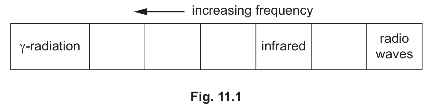
(a) X-rays are placed between ultraviolet and \(\gamma\)-radiation in the electromagnetic spectrum.
The spectrum is ordered by increasing frequency: radio waves → microwaves → infrared → visible → ultraviolet → X-rays → gamma radiation. X-rays have a higher frequency than ultraviolet light but a lower frequency than gamma radiation, so they belong in the gap between those two.

(b)(i) One hospital use of X-rays: imaging bones / detecting fractures (radiography).
X-rays penetrate soft tissue but are absorbed by denser materials such as bone, creating shadow images on a detector.
This makes them ideal for diagnosing broken bones, dental problems, or screening for tumours without the need for surgery.
(b)(ii) Ultrasound is used instead of X-rays because it is non-ionising and therefore harmless to the developing foetus.
X-rays are ionising radiation that can damage DNA and cause mutations, posing a serious risk to rapidly dividing foetal cells.
Ultrasound uses high-frequency sound waves that carry no such risk, making it the safe and preferred choice for prenatal scanning.
(b)(iii) Ultrasound has a frequency above 20 kHz — the upper limit of human hearing.
Humans can hear sounds between approximately 20 Hz and 20 kHz; any frequency above this range is called ultrasound.
Medical ultrasound typically operates in the range of thousands of kHz (MHz), well beyond what the human ear can detect.
(c) Wavelength of \(\gamma\)-radiation:
\(\lambda = \dfrac{v}{f} = \dfrac{3 \times 10^{8}}{6 \times 10^{19}} = 5 \times 10^{-12}\,\textbf{m}\)
This extremely short wavelength (5 picometres) is characteristic of gamma radiation and reflects its very high frequency.
All electromagnetic waves share the same wave equation \(v = f\lambda\), with speed \(3 \times 10^8\,\text{m/s}\) in a vacuum.
(d)(i) … half the nuclei of that isotope in any sample to decay.
Half-life is a fixed, characteristic value for each radioactive isotope and is independent of sample size or external conditions.
It describes the statistical rate of random decay — after each half-life, exactly half of the remaining radioactive nuclei will have decayed.
(d)(ii) Number of cobalt-60 atoms remaining after 31.8 years = 25.
\(31.8 \div 5.3 = 6\) half-lives.
\(1600 \times \left(\tfrac{1}{2}\right)^6 = 1600 \div 64 = 25\,\text{atoms}\).
Each half-life halves the count: 1600 → 800 → 400 → 200 → 100 → 50 → 25.