Breathing System – Key Structures to Identify
🌬️ Main Parts to Identify
- Lungs
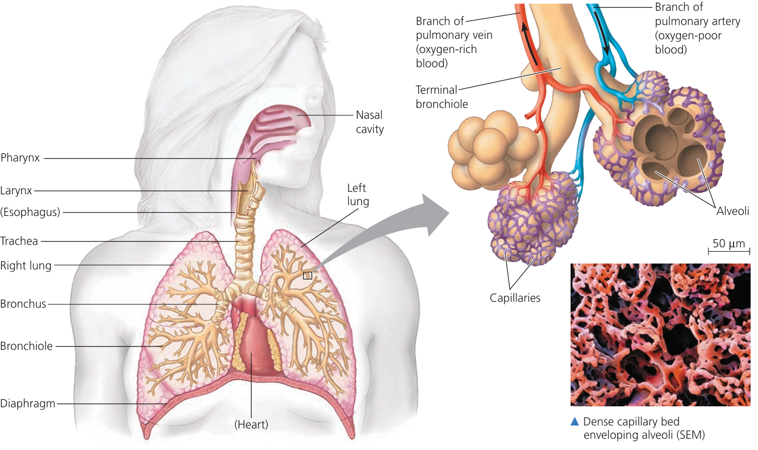
Two large spongy organs in the chest.
Filled with bronchi, bronchioles, and alveoli.
Function: Main site of gas exchange. - Diaphragm
Dome-shaped sheet of muscle under the lungs.
Contracts → flattens → increases chest volume (inhalation).
Relaxes → curves upward → decreases volume (exhalation). - Ribs
Bones forming rib cage around lungs and heart.
Protect lungs + assist in breathing (move up and out during inhalation). - Intercostal Muscles
Muscles between ribs.
Work with diaphragm to expand/contract chest.
External intercostals: help inhale.
Internal intercostals: help forceful exhale. - Larynx (Voice Box)
Located at top of trachea.
Contains vocal cords. - Trachea (Windpipe)
Tube carrying air from throat → bronchi.
Lined with cartilage rings (to keep it open). - Bronchi
Two branches from trachea → each lung.
Carry air into lungs. - Bronchioles
Smaller branches of bronchi inside lungs.
Spread air to alveoli. - Alveoli
Tiny air sacs at ends of bronchioles.
Thin walls → surrounded by capillaries.
Site of gas exchange (O₂ in, CO₂ out). - Associated Capillaries
Fine blood vessels covering alveoli.
Allow diffusion of gases between blood and air.
Oxygen → blood, CO₂ → alveoli.
📊 Summary Table
| Structure | Location | Function | Diagram Feature |
|---|---|---|---|
| Lungs | Chest cavity | Gas exchange | Two spongy sacs |
| Diaphragm | Below lungs | Breathing movements | Dome-shaped muscle |
| Ribs | Surround chest | Protection + movement | Bony arcs |
| Intercostal muscles | Between ribs | Expand/contract chest | Thin bands |
| Larynx | Top of trachea | Voice + air passage | Bulge at throat |
| Trachea | Neck → chest | Air passage | Tube with cartilage rings |
| Bronchi | From trachea → lungs | Air conduction | Two branches |
| Bronchioles | Inside lungs | Air distribution | Fine tubes |
| Alveoli | Ends of bronchioles | Gas exchange | Tiny sacs |
| Capillaries | Around alveoli | Gas diffusion | Web around sacs |
⚡ Quick Recap
Big picture: Air enters via trachea → bronchi → bronchioles → alveoli → capillaries.
Muscles involved: Diaphragm + intercostal muscles.
Protective framework: Ribs.
Special structures: Larynx = voice box.
Exchange site: Alveoli + capillaries (like grapes wrapped in a net).
Investigating Inspired vs Expired Air with Limewater
🌱 Introduction
We can compare inspired (inhaled) air and expired (exhaled) air to see how their gas composition changes in the lungs.
The simplest school experiment uses limewater as a test for carbon dioxide (CO₂).
🧪 Principle of the Test
- Limewater = dilute calcium hydroxide solution.
- CO₂ present → limewater turns milky/cloudy (due to calcium carbonate formation).
Equation:
\[
\text{Ca(OH)}_{2} \,(aq) + \text{CO}_{2} \,(g) \;\rightarrow\; \text{CaCO}_{3} \,(s) + \text{H}_{2}O \,(l)
\]
🔧 Apparatus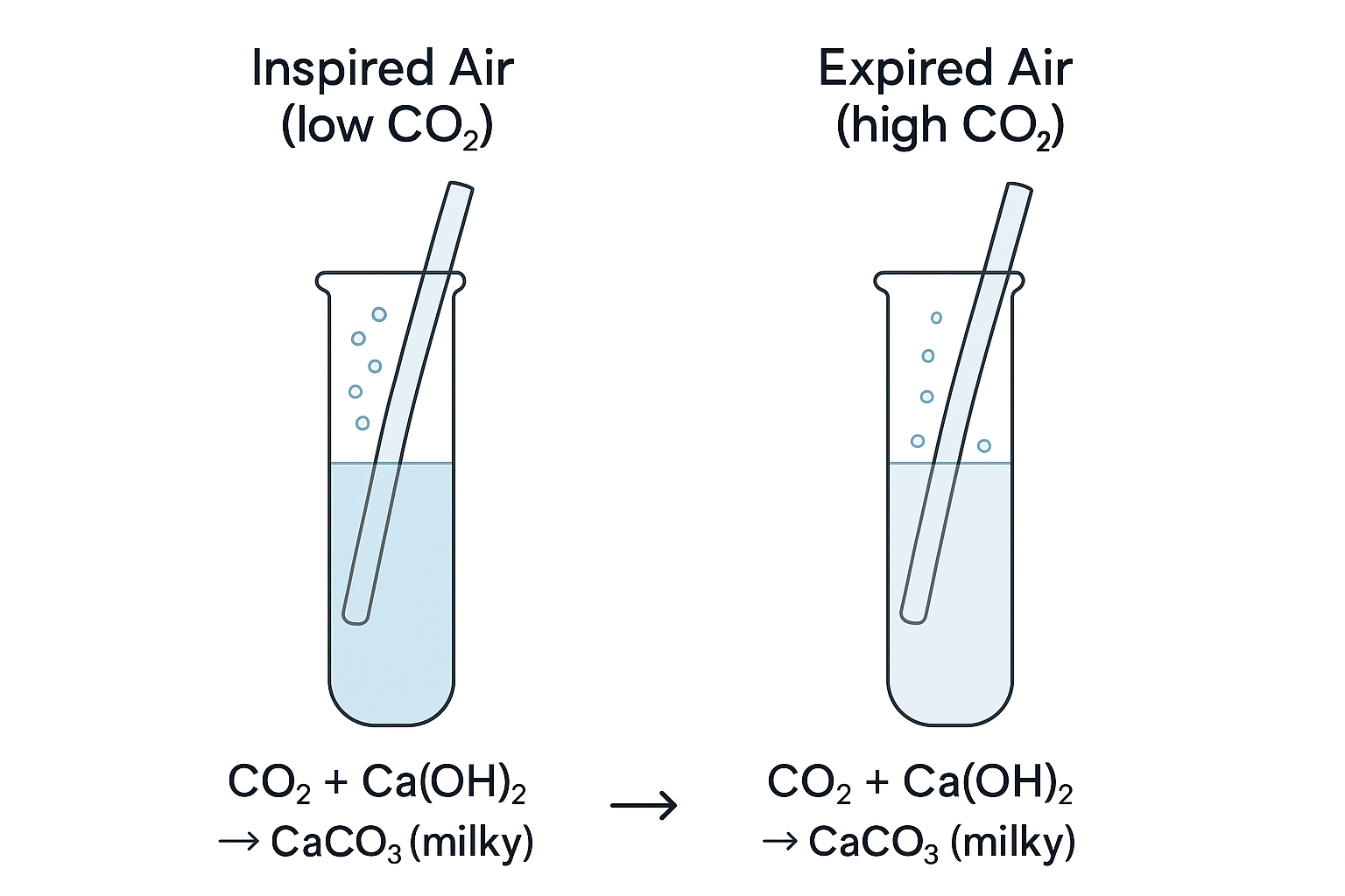
- Two test tubes or beakers with equal amounts of fresh limewater
- Drinking straws / tubing (clean, separate for each test)
- Clamp stand / rack
🧭 Method
- Fill two containers with equal volumes of limewater.
- Inspired air (control for room air):
Bubble normal room air through limewater (using a syringe or pump, or simply blowing in gently with a straw).
Note how long it takes to turn milky. - Expired air:
Exhale gently into the second limewater container using a clean straw.
Observe how quickly it turns milky. - Compare the results between the two.
- Repeat to improve reliability.
🎯 Observations
- Room/inspired air: Limewater turns milky slowly or very faintly (low CO₂).
- Expired air: Limewater turns milky much faster (high CO₂).
🧠 Explanation
In the alveoli:
- Oxygen diffuses into blood.
- Carbon dioxide diffuses out of blood.
So expired air has:
- Less oxygen
- More carbon dioxide
- More water vapour (air gets humidified in the lungs)
📊 Summary Table
| Gas | Inspired (inhaled) air | Expired (exhaled) air | Limewater test |
|---|---|---|---|
| Oxygen | ~21% | ~16% | Not tested here |
| Carbon dioxide | ~0.04% | ~4% | Limewater turns milky faster |
| Water vapour | Variable | Higher | Not tested here |
📝 Conclusion
Expired air has more CO₂ than inspired air.
This is proven by the limewater test: it becomes milky much quicker when bubbled with expired air.
The difference is due to gas exchange during respiration.
⚡ Quick Recap
Test used: limewater → CO₂ turns it milky.
Expired air → limewater cloudy fast (more CO₂).
Inspired air → limewater slower change (less CO₂).
Explains: O₂ taken in, CO₂ + H₂O breathed out.
Effects of Physical Activity on Breathing
🌱 Introduction
During physical activity, muscles need more energy for contraction. This energy comes from increased respiration, which uses more oxygen and produces more carbon dioxide.
To meet this demand, our breathing pattern changes.
🔬 What to Investigate
We can measure:
- Breathing rate → number of breaths per minute.
- Breathing depth → how much air is taken in/out per breath (tidal volume).
📊 Method (School Investigation Idea)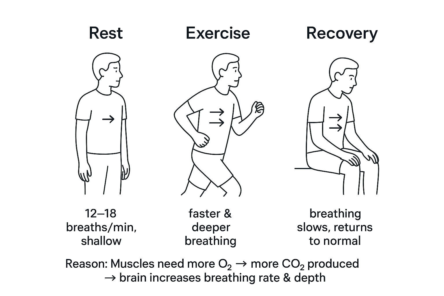
- Count breaths per minute at rest (normal breathing).
- Perform moderate exercise (e.g. jogging on the spot for 1–2 min).
- Immediately count breaths per minute again.
- Observe the depth of breathing (shallow vs deep chest movements).
- Compare results.
🎯 Observations
- At rest:
Breathing rate = slower (about 12–18 breaths/min).
Breathing depth = shallow/normal. - During exercise:
Breathing rate = faster (can double or triple).
Breathing depth = much deeper (lungs take in more air per breath). - After exercise:
Breathing rate stays high for a short time, then gradually returns to normal.
🧠 Biological Explanation
- Exercise increases muscle activity → more ATP needed.
- Respiration rate rises, consuming more O₂ and releasing more CO₂.
- Brain (medulla) detects ↑ CO₂ levels in blood → sends signals to breathing muscles.
- Intercostal muscles + diaphragm contract faster and stronger → breathing becomes faster and deeper.
This ensures:
- More O₂ uptake into blood.
- Faster removal of CO₂.
📊 Summary Table
| Condition | Breathing Rate | Breathing Depth | Reason |
|---|---|---|---|
| Rest | Slow (12–18/min) | Shallow/normal | Low O₂ demand |
| Exercise | Fast (↑↑) | Deep (↑ tidal volume) | High O₂ demand, more CO₂ removal |
| Recovery | Gradually decreases | Deep at first, then normal | Body repays oxygen debt |
⚡ Quick Recap
Physical activity → faster + deeper breathing.
Reason = more O₂ needed, more CO₂ produced.
Controlled by brain’s breathing centre responding to CO₂ levels.
Recovery period = “oxygen debt repayment.”
👉 Memory hook: “Run more → breathe more (fast + deep).”
Physical Activity and Breathing Control
📌 Introduction
During exercise, breathing becomes faster and deeper. This is controlled automatically by the brain in response to changes in the blood.
🌬️ Step-by-Step Link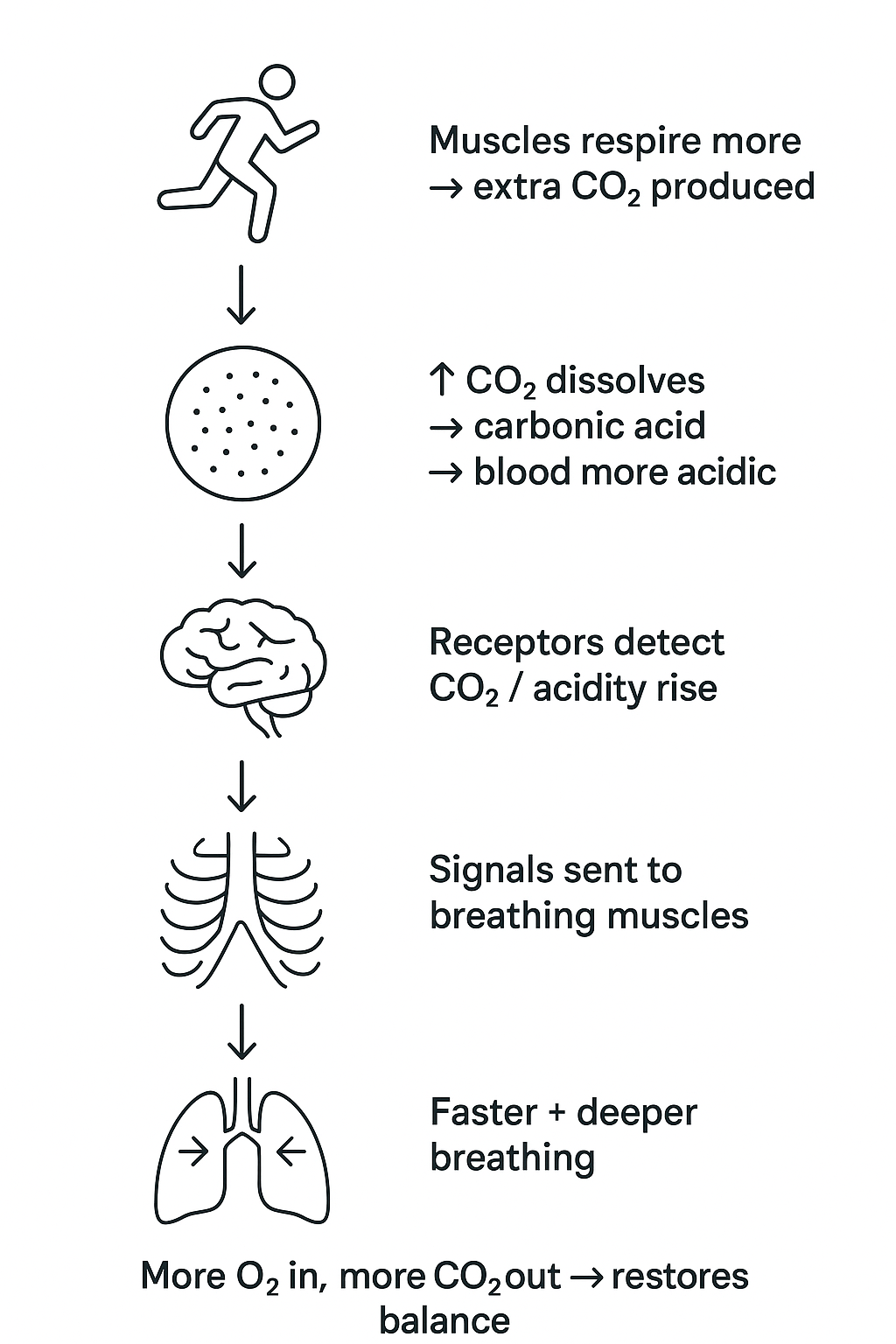
- Exercise increases respiration
Muscles respire more to release energy for contraction.
This produces extra CO₂ as a waste product. - CO₂ concentration rises in blood
Carbon dioxide dissolves in blood → forms carbonic acid.
Blood becomes slightly more acidic. - Detection by brain
Special receptors in the brain (medulla) sense the increase in CO₂ / acidity. - Response sent to breathing muscles
Brain sends nerve signals to the diaphragm and intercostal muscles. - Breathing rate and depth increase
Faster breathing = more breaths per minute.
Deeper breathing = greater volume of air per breath. - Effect
More oxygen enters alveoli → diffuses into blood.
More carbon dioxide is removed from blood → exhaled.
Restores normal gas levels in blood.
📊 Summary Table
| Stage | Event | Result |
|---|---|---|
| Exercise | ↑ Respiration in muscles | ↑ CO₂ produced |
| Blood | ↑ CO₂ concentration | Blood more acidic |
| Brain detects | Medulla senses CO₂ | Sends signals to muscles |
| Breathing response | Faster + deeper | ↑ O₂ in, ↑ CO₂ out |
⚡ Quick Recap
Exercise → more CO₂ in blood.
Brain detects CO₂ rise.
Signals → diaphragm + intercostals.
Breathing becomes faster + deeper.
Ensures: more O₂ supply + CO₂ removal.
👉 Memory hook: “CO₂ tells the brain → breathe more and breathe deeper.”