Plant Cell vs Animal Cell
📌 Introduction
- All living things are made of cells.
- Cells contain organelles that carry out life processes.
- Plant and animal cells share many features but also have key differences.
🔑 Common Structures (found in both) 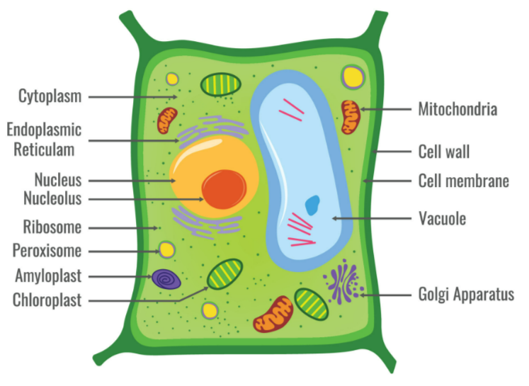
- Cell Membrane
Thin, partially permeable boundary around cytoplasm.
Controls entry & exit of substances. Maintains cell contents. - Cytoplasm
Jelly-like fluid (90% water, with salts, sugars, proteins, enzymes).
Site of many chemical reactions. Contains organelles (mitochondria, ribosomes, etc.). - Nucleus
Rounded structure enclosed by nuclear membrane.
Contains DNA (chromosomes). Controls cell activities, enzyme production, and cell division. - Mitochondria
“Powerhouse of the cell”.
Site of aerobic respiration → energy (ATP) release.
- Ribosomes
Very small organelles.
Site of protein synthesis.
🌿 Extra Features in Plant Cells
- Cell Wall
Made of cellulose (non-living, freely permeable).
Provides shape & support. Prevents bursting. - Chloroplasts
Contain chlorophyll → trap light for photosynthesis.
Absent in animal cells. - Large Permanent Vacuole
Fluid-filled sac with cell sap (sugars, salts, pigments).
Maintains firmness (turgor pressure).
Note: Animal cells may have small, temporary vacuoles only.
📊 Comparison Table
| Feature | Plant Cell | Animal Cell |
|---|---|---|
| Cell wall | Present (cellulose) | Absent |
| Cell membrane | Present | Present |
| Nucleus | Present | Present |
| Cytoplasm | Present | Present |
| Mitochondria | Present | Present |
| Ribosomes | Present | Present |
| Chloroplasts | Present (in green cells) | Absent |
| Vacuole | Large, permanent (cell sap) | Small/temporary, sometimes absent |
⚡ Quick Recap
Both have: cell membrane, cytoplasm, nucleus, mitochondria, ribosomes.
Plant extras = Cell wall + Chloroplast + Large vacuole.
Cell wall = support, Chloroplast = photosynthesis, Vacuole = firmness.
Animal cells → more flexible, no photosynthesis, no cellulose wall.
Permanent vacuole, Photosynthetic chloroplast, Protective cell wall.
Structure of a Bacterial Cell
📌 Introduction
- Bacteria are prokaryotic cells (simple cells without a true nucleus or membrane-bound organelles).
- Much smaller than plant/animal cells.
- Still perform all life processes.
🔑 Main Structures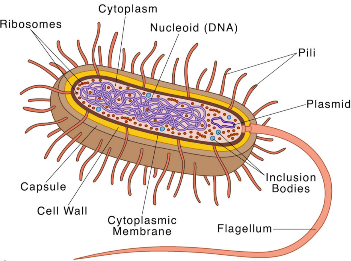
- Cell Wall
Provides shape & protection.
Made of peptidoglycan (not cellulose like plants).
Prevents cell from bursting in dilute solutions. - Cell Membrane
Lies just inside the wall.
Controls entry and exit of substances.
Site of some respiration enzymes (since no mitochondria). - Cytoplasm
Jelly-like fluid containing enzymes for metabolic reactions.
No membrane-bound organelles. - Ribosomes
Small, scattered in cytoplasm.
Site of protein synthesis.
Simpler (70S type) than in eukaryotes. - Circular DNA
Single, long circular molecule of DNA.
Lies free in cytoplasm (no nucleus).
Controls cell activities. - Plasmids
Small extra loops of DNA.
Carry additional genes (e.g. antibiotic resistance).
Can be passed between bacteria.
📊 Summary Table
| Structure | Description | Function |
|---|---|---|
| Cell wall | Peptidoglycan layer | Support & protection |
| Cell membrane | Thin, partially permeable | Controls exchange of substances |
| Cytoplasm | Jelly-like fluid | Site of reactions |
| Ribosomes | Small (70S) | Protein synthesis |
| Circular DNA | Large loop, free in cytoplasm | Main genetic material |
| Plasmids | Small DNA circles | Extra genes (e.g. resistance) |
⚡ Quick Recap
Bacteria = prokaryotes (no nucleus, no mitochondria).
Genetic material = circular DNA + plasmids.
Proteins made on ribosomes (70S).
Energy from enzymes in cell membrane (no mitochondria).
Wall = peptidoglycan, not cellulose.
Identifying Cell Structures (Plant, Animal, Bacteria)
🌱 Plant Cell (typical palisade cell)
Key structures to identify in diagrams/images:
- Cell wall → thick outline (rigid box shape).
- Cell membrane → thin line just inside wall (hard to see separately).
- Cytoplasm → thin layer around vacuole.
- Nucleus → dark, circular/oval body.
- Chloroplasts → green oval structures (often near edges).
- Mitochondria → tiny bean-like shapes (hard in light microscope).
- Ribosomes → too small to see in light microscope, only in diagrams.
- Large vacuole → clear/empty-looking space in centre (filled with cell sap).
🐾 Animal Cell (typical cheek/liver cell)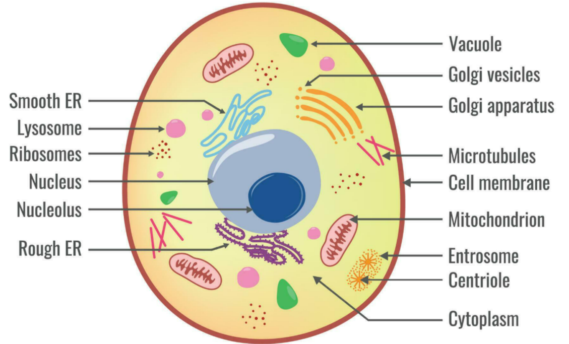
Key structures to identify:
- Cell membrane → thin boundary, outer layer (no cell wall).
- Cytoplasm → grainy liquid filling the cell.
- Nucleus → large, central/dark-stained circle.
- Mitochondria → small rod-like dots (shown in diagrams).
- Ribosomes → shown as tiny dots (not visible in simple light micrographs).
- No chloroplasts, no large vacuole, no cell wall (key difference from plants).
🦠 Bacterial Cell (prokaryote)
Key structures to identify:
- Cell wall → outer layer (not cellulose).
- Cell membrane → inside the wall.
- Cytoplasm → grainy filling (no nucleus).
- Ribosomes → scattered, very small.
- Circular DNA → large loop in cytoplasm (no nuclear envelope).
- Plasmids → small extra loops of DNA (shown as tiny circles).
- No mitochondria, no chloroplasts, no true nucleus.
📊 At-a-Glance ID Guide
| Structure | Plant | Animal | Bacteria |
|---|---|---|---|
| Cell wall | ✓ | ✘ | ✓ (peptidoglycan) |
| Cell membrane | ✓ | ✓ | ✓ |
| Cytoplasm | ✓ | ✓ | ✓ |
| Nucleus | ✓ | ✓ | ✘ (DNA free in cytoplasm) |
| Chloroplasts | ✓ (green cells only) | ✘ | ✘ |
| Mitochondria | ✓ | ✓ | ✘ |
| Ribosomes | ✓ | ✓ | ✓ (smaller) |
| Vacuole | Large, permanent | Small/temporary | ✘ |
| Circular DNA | ✘ | ✘ | ✓ |
| Plasmids | ✘ | ✘ | ✓ |
⚡ Quick Recap
Plant cells → box-shaped, cell wall, chloroplasts, big vacuole.
Animal cells → round/irregular, no wall, no chloroplast, only small vacuoles.
Bacteria → tiny, no nucleus, DNA loop + plasmids, no mitochondria.
Cell Division and Formation of New Cells
📌 Key Point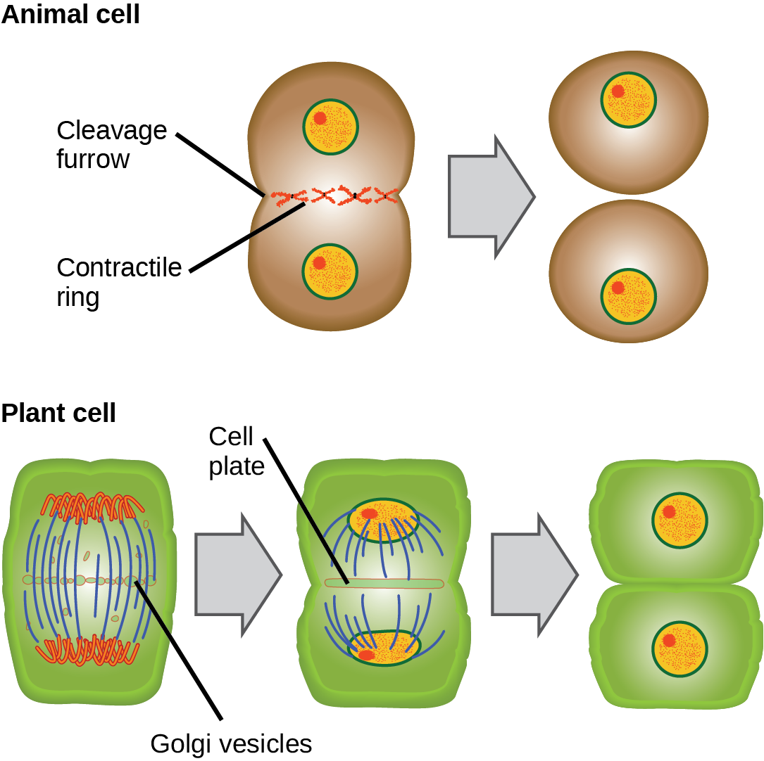
- New cells are always produced by the division of existing cells.
- This principle is part of the cell theory.
🔑 Cell division allows:
- Growth → increase in number of cells.
- Repair → replacement of damaged or dead cells.
- Reproduction → formation of gametes or new organisms (depending on type of division).
Methods:
- Mitosis → produces genetically identical cells (growth, repair).
- Meiosis → produces gametes (variation).
⚡ Quick Recap
Cells don’t appear from nowhere.
All cells come from pre-existing cells by division.
Division = essential for growth, repair, and reproduction.
Levels of Organisation
📌 Introduction
- Living organisms are built in a hierarchical order.
- Smallest unit = cell, largest unit = organism.
- Each level is more complex than the previous one.
🔑 Key Terms & Examples
1. Cell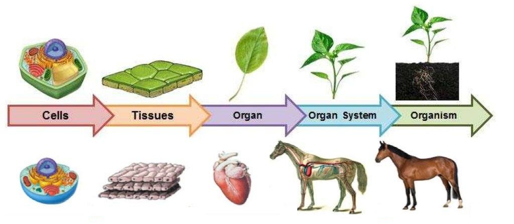
- The basic structural & functional unit of life.
- Smallest part of an organism that can work independently.
- Examples:
- Plant: Palisade mesophyll cell (photosynthesis).
- Animal: Red blood cell (oxygen transport).
2. Tissue
- A group of similar cells working together to perform a particular function.
- Examples:
- Plant: Xylem tissue (transports water).
- Animal: Muscle tissue (contracts for movement).
3. Organ
- A structure made of different tissues grouped together for a specific function.
- Examples:
- Plant: Leaf (photosynthesis).
- Animal: Stomach (digestion).
4. Organ System
- A group of organs working together to carry out a major body function.
- Examples:
- Plant: Shoot system (stem + leaves + buds → supports plant + photosynthesis).
- Animal: Circulatory system (heart + blood vessels → transport of blood).
5. Organism
- A living individual made up of organ systems that work together.
- Examples:
- Plant: Sunflower.
- Animal: Human being.
📊 Summary Table – Levels of Organisation
| Level | Definition | Example (Plant) | Example (Animal) |
|---|---|---|---|
| Cell | Basic unit of life | Root hair cell | Red blood cell |
| Tissue | Group of similar cells | Xylem tissue | Muscle tissue |
| Organ | Made of tissues with a function | Leaf | Stomach |
| Organ System | Group of organs | Shoot system | Circulatory system |
| Organism | Complete living thing | Sunflower | Human |
⚡ Quick Recap
Cell → Tissue → Organ → Organ System → Organism (CTOSO).
Examples:
Plant: Root hair cell → Xylem → Leaf → Shoot system → Sunflower.
Animal: RBC → Muscle → Stomach → Digestive system → Human.