CIE iGCSE Co-ordinated Sciences-B2.2 Size of specimens- Study Notes- New Syllabus
CIE iGCSE Co-ordinated Sciences-B2.2 Size of specimens – Study Notes
CIE iGCSE Co-ordinated Sciences-B2.2 Size of specimens – Study Notes -CIE iGCSE Co-ordinated Sciences – per latest Syllabus.
Key Concepts:
- State and use the formula:
magnification = Image Size / Actual Size - Calculate magnification and size of biological specimens using millimetres as units
- Convert measurements between millimetres (mm) and micrometres (µm)
CIE iGCSE Co-Ordinated Sciences-Concise Summary Notes- All Topics
Size of Specimens & Magnification
📌 Why we need microscopes
- Most cells are too small to be seen with naked eye.
- Hand lens (up to ×20) → not enough to see cell detail.
- Light microscope → magnifies up to ×1500 (school ones usually ×400).
🧾 Structure of a Light Microscope
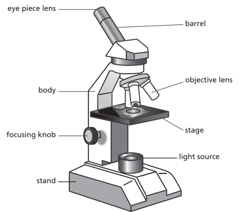
- Eyepiece lens: usually ×10.
- Objective lenses: ×4, ×10, ×40 (rotating nosepiece).
- Total magnification = eyepiece × objective.
- Eg: ×10 eyepiece × ×40 objective = ×400.
- Light passes through specimen → objective lens → eyepiece → magnified image.
- Coarse & fine focus knobs → sharpen image.
📌 Types of microscope slides
- Temporary slide → quick to prepare, dries out fast, not long-lasting.
- Permanent slide → specimen dehydrated + fixed in resin (e.g. Canada Balsam), lasts for years.
- Coverslip → keeps specimen in place, reduces dehydration, protects lens.
🔑 Formula for Magnification
\[ \text{Magnification (M)} = \frac{\text{Image size}}{\text{Actual size}} \]
\[ \text{Actual size} = \frac{\text{Image size}}{\text{Magnification (M)}} \]
Always make sure units are the same (mm ↔ µm ↔ nm).
- 1 cm = 10 mm
- 1 mm = 1000 µm
📊 Example
Q. A plant cell drawing is measured as 20 mm across.
Magnification used = ×400.
Actual size = 20 mm ÷ 400 = 0.05 mm = 50 µm
So, the real cell is 50 micrometres wide.
⚡ Quick Recap
Total magnification = eyepiece × objective.
Units must match.
Light microscope: up to ×1500 (school: ×400).
Temporary vs permanent slides: quick but dry out vs long-lasting
Calculating Magnification & Size (in mm)
🔑 Formula
\[ \text{Magnification (M)} = \frac{\text{Image size}}{\text{Actual size}} \]
\[ \text{Actual size} = \frac{\text{Image size}}{\text{Magnification (M)}} \]
Both image size and actual size must be in mm before calculation.
🔄 Unit Conversions (for mm standard)
- 1 cm = 10 mm
- 1 mm = 1000 µm
- 1 µm = 0.001 mm
So if given in µm, divide by 1000 to convert → mm.
🧮 Examples
Example 1
A diagram of a bacterium is measured as 25 mm.
Real bacterium is 0.005 mm long.
M = 25 ÷ 0.005 = 5000
Magnification = ×5000
Example 2
A drawing of a plant cell is 40 mm across.
Magnification = ×800.
Actual size = 40 ÷ 800 = 0.05 mm
Actual size = 0.05 mm (50 µm)
Example 3
Observed image of a red blood cell = 70 mm.
Real RBC = 0.007 mm.
M = 70 ÷ 0.007 = 10,000
Magnification = ×10,000
⚡ Quick Recap (Millimetre Method)
Always convert → mm before calculation.
Remember: 1 mm = 1000 µm
Quote units in final answer (mm or µm if converted).
Conversion between Millimetres (mm) and Micrometres (µm)
📌 Relationship
- 1 mm = 1000 µm
- 1 µm = 0.001 mm
✨ How to Convert
1. mm → µm
Multiply by 1000
Value in µm = Value in mm × 1000
Example:
0.2 mm = 0.2 × 1000 = 200 µm
2. µm → mm
Divide by 1000
Value in mm = Value in µm ÷ 1000
Example:
2500 µm = 2500 ÷ 1000 = 2.5 mm
🧮 Practice Examples
- 1.5 mm = 1.5 × 1000 = 1500 µm
- 50 µm = 50 ÷ 1000 = 0.05 mm
- 0.03 mm = 0.03 × 1000 = 30 µm
- 12000 µm = 12000 ÷ 1000 = 12 mm
⚡ Quick Recap
Multiply by 1000 → mm to µm
Divide by 1000 → µm to mm
Always keep units clear in the final answer!