Identifying Structures of the Mammalian Heart
📌 Introduction
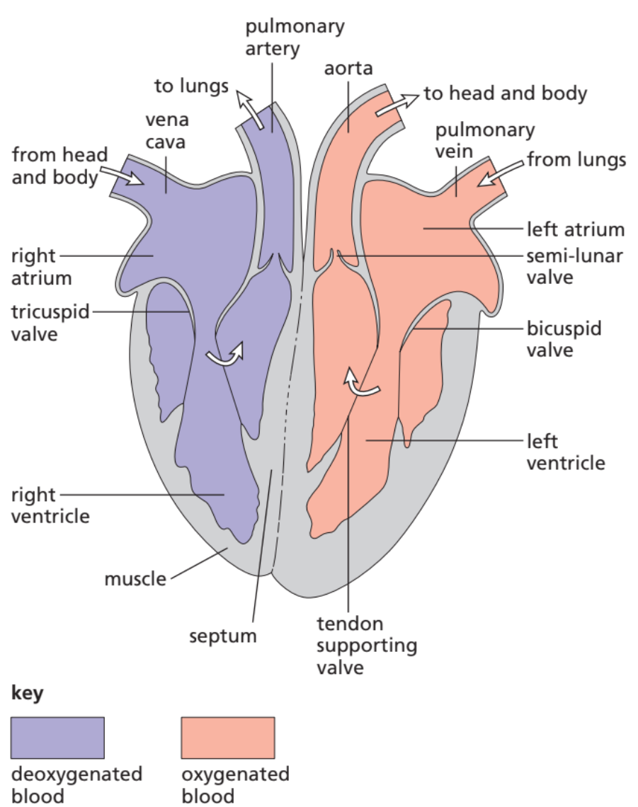
Remember: the heart is drawn as if you are facing the person, so left side of the diagram = right side of the heart.
🔎 Key Structures to Identify
1. Muscular Wall (Myocardium)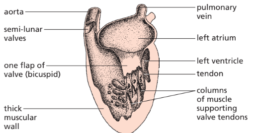
- Thickest around the left ventricle (pumps blood to the whole body).
- Thinner around the right ventricle (pumps blood only to lungs).
- Function: provides strong contractions to pump blood.
2. Septum
- Thick muscular wall dividing the left and right sides of the heart.
- Prevents mixing of oxygenated and deoxygenated blood.
3. Ventricles (Lower Chambers)
- Right ventricle → pumps deoxygenated blood to lungs via pulmonary artery.
- Left ventricle → pumps oxygenated blood to body via aorta.
- Ventricles have thicker walls than atria (because they pump blood further).
4. Atria (Upper Chambers)
- Right atrium → receives blood from body via vena cava.
- Left atrium → receives blood from lungs via pulmonary veins.
- Atria have thinner walls, as they only pump into ventricles.
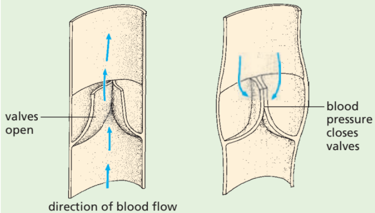
5. One-Way Valves
- Ensure blood flows in one direction only.
- Atrioventricular valves (bicuspid & tricuspid) → between atria and ventricles.
- Semilunar valves → at exits of ventricles (aorta & pulmonary artery).
6. Coronary Arteries
- Tiny arteries on the surface of the heart.
- Supply oxygen and glucose directly to the heart muscle.
- If blocked → causes heart attack (coronary heart disease).
📝 Summary Table
| Structure | Location | Function |
|---|---|---|
| Muscular wall | Around chambers | Pumps blood by contraction |
| Septum | Middle of heart | Separates left & right sides |
| Right ventricle | Lower right | Pumps deoxygenated blood to lungs |
| Left ventricle | Lower left | Pumps oxygenated blood to body |
| Right atrium | Upper right | Receives blood from body |
| Left atrium | Upper left | Receives blood from lungs |
| One-way valves | Between chambers & arteries | Prevent backflow |
| Coronary arteries | On heart surface | Supply heart muscle with oxygen/glucose |
⚡ Quick Recap
Atria = receive blood, ventricles = pump blood.
Left ventricle wall is thicker (pumps to whole body).
Septum prevents blood mixing.
Valves → keep flow one-way.
Coronary arteries feed the heart itself.
Blood Vessels – Direction of Flow
📌 Key Point
- Arteries → carry blood away from the heart.
- Veins → carry blood back to the heart.
🔎 Explanation
The heart = pump that pushes blood around the body.
- When ventricles contract, blood is forced out through arteries.
- After travelling through capillaries in tissues, blood returns via veins into the atria.
📝 Summary Table
| Vessel | Direction of Blood Flow | Example |
|---|---|---|
| Arteries | Away from heart | Aorta, Pulmonary artery |
| Veins | Toward the heart | Vena cava, Pulmonary vein |
⚡ Quick Recap
➡️ Arteries = Away
⬅️ Veins = back to heart
(Easy trick: A = Away, V = Visit back to heart)
Monitoring Heart Activity
📌 Key Idea
The activity of the heart can be monitored using different methods:
🔎 Methods
1. ECG (Electrocardiogram)
- Records the electrical activity of the heart.
- Electrodes attached to the skin detect impulses when atria & ventricles contract.
- Produces a graph trace (ECG) showing rhythm, strength, and regularity.
- Useful to detect abnormal rhythms, heart attacks, or heart disease.
2. Pulse Rate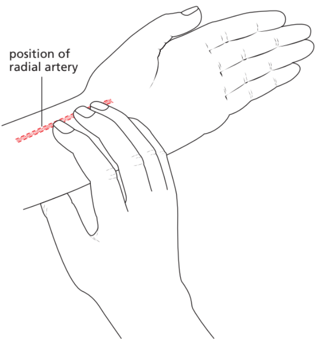
- Pulse = pressure wave caused by contraction of ventricles pushing blood into arteries.
- Felt at wrist or neck.
- Shows heart rate (beats per minute) and rhythm.
3. Listening to Valve Sounds (Stethoscope)
- A stethoscope amplifies sounds of the atrioventricular and semilunar valves closing.
- “Lub-dub” sound = normal heartbeat.
- Lub → AV valves closing.
- Dub → Semilunar valves closing.
- Helps detect leaky valves or irregular heartbeat.
📝 Summary Table
| Method | What it Detects | Example Use |
|---|---|---|
| ECG | Electrical activity of heart | Heart attack, arrhythmia |
| Pulse rate | Pressure wave in arteries | Measuring heart rate |
| Valve sounds | Valve closure sounds | Detecting murmurs/leaky valves |
⚡ Quick Recap
ECG → electricity of heart
Pulse → beats per minute
Stethoscope → “lub-dub” valve sounds
Coronary Heart Disease (CHD)
📌 Definition
Coronary Heart Disease (CHD) occurs when coronary arteries (the blood vessels supplying the heart muscle with oxygen + glucose) become blocked or narrowed, usually by fatty deposits (plaque).
This reduces blood flow → less oxygen supply → heart muscle may be damaged (angina, heart attack).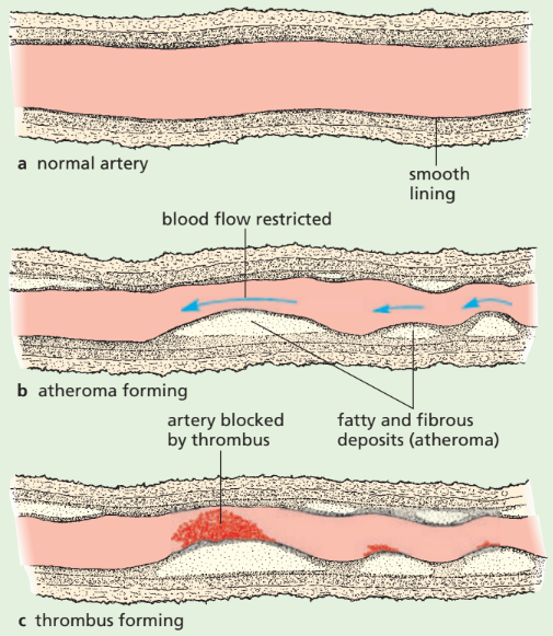
🔬 How it Happens
- Fatty deposits (cholesterol + plaque) build up in coronary arteries.
- Artery walls become narrow and less flexible (atherosclerosis).
- Blood flow to heart muscle reduces → less oxygen and glucose for respiration.
- If completely blocked → heart attack (myocardial infarction).
⚠️ Risk Factors
1. Diet
- High in saturated fats & cholesterol → more plaque formation.
2. Lack of exercise
- Leads to obesity → raises blood pressure & cholesterol.
3. Stress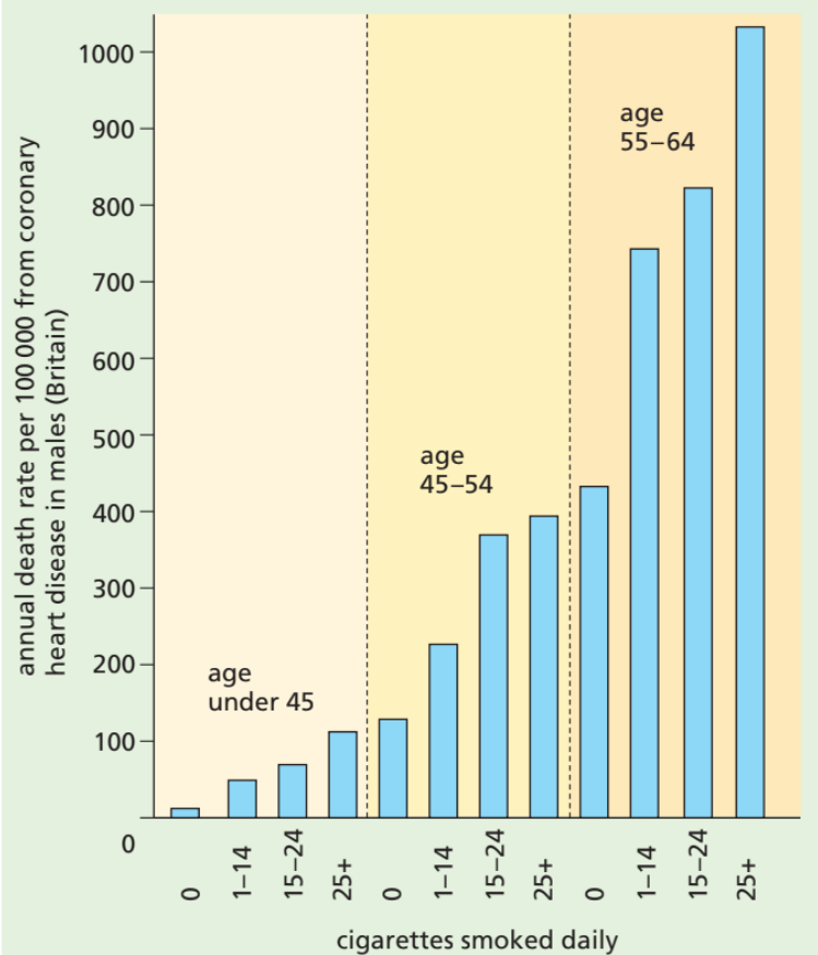
- Increases blood pressure → damages artery walls.
4. Smoking
- Nicotine ↑ blood pressure, damages arteries.
- Carbon monoxide reduces oxygen carrying capacity of blood.
5. Genetic predisposition
- Family history → higher chance of CHD.
6. Age
- Older people more likely to have fatty deposits.
7. Sex
- Men at higher risk (before menopause, women have protective effect of estrogen).
📝 Summary Table
| Risk Factor | Effect |
|---|---|
| Diet (high fat) | More cholesterol → artery blockage |
| Lack of exercise | Obesity, ↑ BP, weaker heart |
| Stress | ↑ BP, artery damage |
| Smoking | Nicotine (↑ BP), CO (less O₂ transport) |
| Genetics | Inherited risk |
| Age | Older = arteries less elastic |
| Sex | Men higher risk than pre-menopausal women |
⚡ Quick Recap
CHD = blockage of coronary arteries → less oxygen → heart attack risk.
Main risks = diet, no exercise, stress, smoking, genetics, age, sex.
Prevent by: healthy diet + regular exercise + no smoking.
Functioning of the Heart
📌 Introduction
The heart works as a pump using rhythmic contraction and relaxation of its muscular walls. Valves ensure one-way flow of blood.
1. Contraction of Atria (Atrial Systole)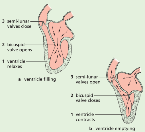
- Both atria contract at the same time.
- Blood is pushed:
- Right atrium → Right ventricle (through tricuspid valve).
- Left atrium → Left ventricle (through bicuspid/mitral valve).
- Valves open because of pressure from atria > ventricles.
2. Contraction of Ventricles (Ventricular Systole)
- Ventricles contract strongly (thicker walls than atria).
- Blood is pumped:
- Right ventricle → Pulmonary artery → Lungs (deoxygenated blood).
- Left ventricle → Aorta → Rest of body (oxygenated blood).
- Atrioventricular (AV) valves close → prevents backflow into atria.

- Semi-lunar (SL) valves open → allow blood out to arteries.
3. Relaxation Phase (Diastole)
- Both atria and ventricles relax.
- Blood flows into atria from:
- Vena cava → Right atrium.
- Pulmonary veins → Left atrium.
- AV valves reopen → cycle repeats.
🔑 Role of Valves
- Atrioventricular valves (tricuspid & bicuspid): Prevent backflow from ventricles → atria.
- Semi-lunar valves (pulmonary & aortic): Prevent backflow from arteries → ventricles.
- Valves open/close due to pressure differences (not by muscles).
📝 Summary Table
| Stage | Action | Valves | Blood Flow |
|---|---|---|---|
| Atrial systole | Atria contract | AV valves open | Atria → Ventricles |
| Ventricular systole | Ventricles contract | AV valves close, SL valves open | Ventricles → Arteries |
| Diastole | Heart relaxes | AV valves reopen | Veins → Atria → Ventricles |
⚡ Quick Recap
Atria contract first → fill ventricles.
Ventricles contract → pump blood out.
Valves = one-way doors → stop backflow.
Cycle = atrial systole → ventricular systole → diastole.