Blood Vessels – Arteries, Veins & Capillaries
📌 Introduction
Blood moves around the body through three main types of vessels:
- Arteries → carry blood away from the heart at high pressure.
- Veins → return blood to the heart at low pressure.
- Capillaries → tiny exchange vessels between blood & body cells.
🔴 Arteries
- Function → carry blood away from heart (mostly oxygenated, except pulmonary artery).
- Wall: very thick, muscular & elastic → withstands and maintains high pressure.
- Lumen: narrow (smaller diameter) → keeps blood under pressure.
- Valves: absent (pressure is already high, prevents backflow).
- Adaptation → Elastic fibres stretch + recoil with each heartbeat → maintains a pressure wave (pulse).
🔵 Veins
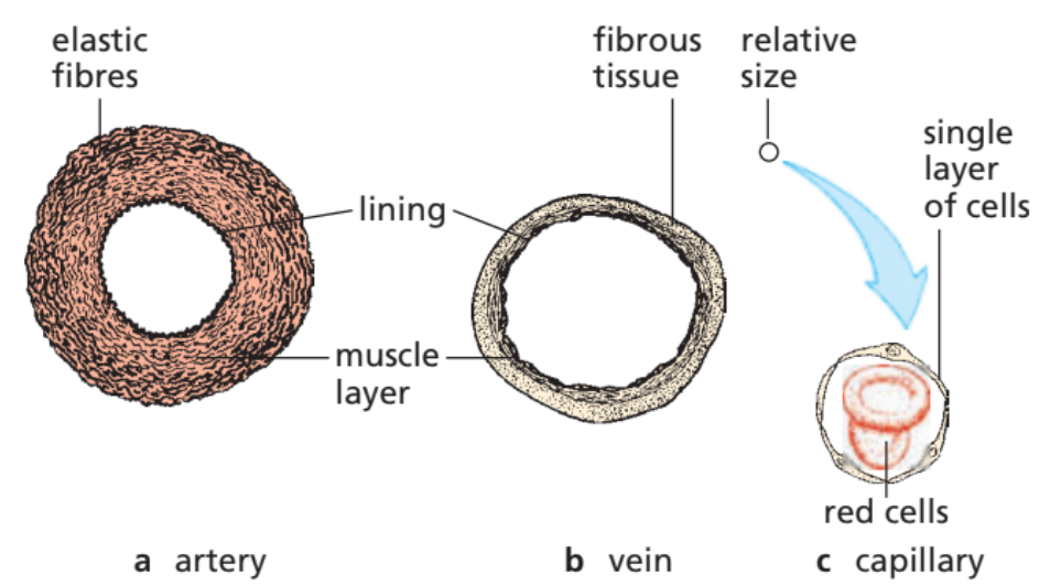
- Function → carry blood back to heart (mostly deoxygenated, except pulmonary vein).
- Wall: thin, with little muscle & elastic tissue → blood pressure is low.
- Lumen: wide → reduces resistance, helps smooth flow.
- Valves: present → prevent backflow (important because pressure is low).
- Adaptation → Movement of body muscles squeezes veins → valves ensure one-way flow toward heart.
⚪ Capillaries
- Function → site of exchange of substances between blood & tissues.
- Wall: one cell thick, permeable → allows diffusion.
- Lumen: extremely narrow (just wide enough for single red blood cells to pass).
- Valves: absent.
- Adaptation → Slow blood flow + thin walls = efficient diffusion of O₂, CO₂, nutrients & wastes.
📊 Summary Table – Comparison
| Feature | Arteries | Veins | Capillaries |
|---|---|---|---|
| Wall thickness | Very thick (muscle + elastic) | Thin, little muscle | One cell thick |
| Lumen size | Narrow | Wide | Very narrow (1 RBC wide) |
| Valves | Absent | Present | Absent |
| Pressure | High | Low | Drops from artery to vein |
| Function | Carry blood away from heart | Carry blood back to heart | Exchange of substances |
⚡ Quick Recap
Arteries = thick walls, narrow lumen, no valves, high pressure.
Veins = thin walls, wide lumen, valves, low pressure.
Capillaries = one cell thick, tiny lumen, site of exchange.
Functions of Capillaries
📌 Introduction
Capillaries are the smallest blood vessels, forming dense networks between arteries and veins. They are the main exchange surface between blood and body cells.
🌱 Main Functions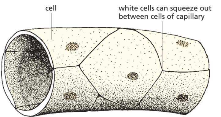
- Exchange of gases
Oxygen diffuses from blood → tissues.
Carbon dioxide diffuses from tissues → blood. - Nutrient delivery
Glucose, amino acids, and other nutrients move from blood → cells. - Removal of wastes
Urea and other metabolic wastes move from cells → blood. - Maintain tissue fluid
Plasma leaks out under pressure → forms tissue fluid, bathing cells. - Immune defense
White blood cells can squeeze through gaps in the wall → fight infection in tissues. - Slow blood flow
Narrow lumen slows RBCs → allows more time for diffusion.
⚡ Quick Recap
Capillaries = exchange vessels
Thin wall (1 cell thick) → fast diffusion.
Narrow lumen → slows blood, 1 RBC at a time.
Functions → O₂ & nutrient supply, CO₂ & waste removal, WBC escape, tissue fluid formation.
Structure of Arteries vs Veins & Blood Pressure
📌 Introduction
Arteries and veins have different structures because the pressure of blood in them is not the same.
- Arteries → carry blood away from heart at high pressure.
- Veins → carry blood back to heart at low pressure.
🌱 Arteries (High Pressure Adaptations)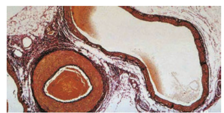
- Thick muscular + elastic walls → resist the high pressure and prevent bursting.
- Elastic fibres → allow walls to stretch with each heartbeat (pulse) and recoil to maintain pressure.
- Narrow lumen → maintains pressure as blood flows through.
- No valves (except at heart exits) → high pressure already prevents backflow.
🌊 Veins (Low Pressure Adaptations)
- Thinner walls → less muscle and elastic tissue (not needed for high pressure).
- Large lumen → reduces resistance and helps easy flow of blood at low pressure.
- Valves present → prevent backflow since pressure is low.
- Assisted by skeletal muscles → contraction squeezes veins and pushes blood back to the heart.
📝 Summary Table
| Feature | Arteries (High Pressure) | Veins (Low Pressure) |
|---|---|---|
| Wall thickness | Very thick, muscular, elastic | Thin, less muscle/elastic |
| Lumen size | Narrow | Wide |
| Valves | Absent (except near heart) | Present (to stop backflow) |
| Function link | Withstand surges of pressure & keep flow continuous | Aid return of low-pressure blood to heart |
⚡ Quick Recap
Arteries → thick, elastic walls + narrow lumen → withstand & maintain high pressure.
Veins → thin walls + wide lumen + valves → transport blood at low pressure without backflow.
Main Blood Vessels – Heart & Lungs
📌 Introduction
The heart has its own major blood vessels that carry blood to and from the body and to and from the lungs. Recognising them in diagrams is a must-know skill for exams.
(a) Blood Vessels of the Heart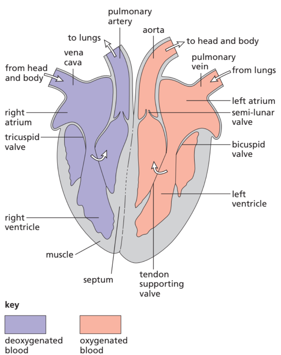
Vena Cava
- Main vein of the body.
- Brings deoxygenated blood from the body → right atrium.
- Two branches:
– Superior vena cava (from head & arms).
– Inferior vena cava (from abdomen & legs).
Aorta
- Largest artery.
- Carries oxygenated blood from left ventricle → rest of the body.
- Very thick wall to withstand high pressure.
Pulmonary Artery
- Carries deoxygenated blood from right ventricle → lungs.
- Only artery in the body that carries deoxygenated blood.
Pulmonary Vein
- Brings oxygenated blood from lungs → left atrium.
- Only vein that carries oxygenated blood.
(b) Blood Vessels of the Lungs
- Pulmonary Artery → takes deoxygenated blood from heart → lungs for oxygenation.
- Pulmonary Vein → brings oxygenated blood back to the heart.
📝 Summary Table
| Organ | Vessel | Type of Blood | Direction |
|---|---|---|---|
| Heart | Vena Cava | Deoxygenated | Body → Heart |
| Heart | Aorta | Oxygenated | Heart → Body |
| Heart | Pulmonary Artery | Deoxygenated | Heart → Lungs |
| Heart | Pulmonary Vein | Oxygenated | Lungs → Heart |
| Lungs | Pulmonary Artery | Deoxygenated | Heart → Lungs |
| Lungs | Pulmonary Vein | Oxygenated | Lungs → Heart |
⚡ Quick Recap
Vena Cava = body → heart (deoxy).
Aorta = heart → body (oxy).
Pulmonary Artery = heart → lungs (deoxy).
Pulmonary Vein = lungs → heart (oxy).
👉 Easy Trick:
“Artery Away, Vein Visit” → arteries carry blood away from heart, veins visit the heart. (Exception: pulmonary vessels differ in oxygen content, but rule about direction is always true!)