Components of Blood
📌 Introduction
Blood is a connective tissue that transports substances, protects against disease, and helps maintain homeostasis. It is made of cells (RBCs, WBCs, platelets) suspended in a liquid medium (plasma).
1. Red Blood Cells (RBCs / Erythrocytes)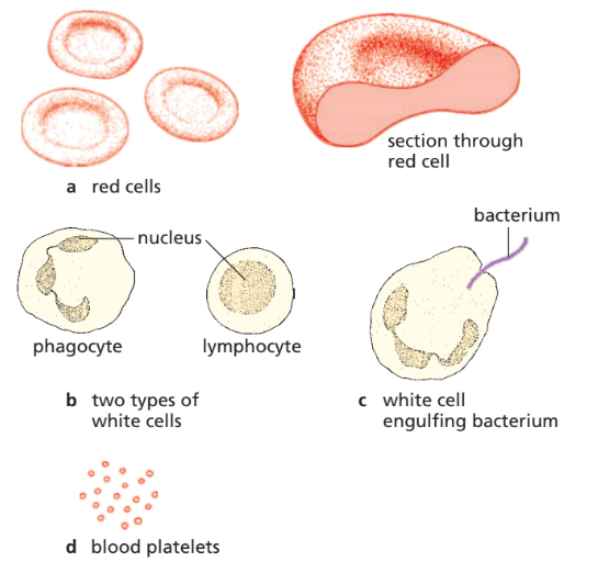
- Structure: Small, biconcave discs (↑ surface area), no nucleus (more space for haemoglobin), flexible to pass through capillaries.
- Function: Transport oxygen (via haemoglobin → oxyhaemoglobin) and small amount of CO₂ (majority in plasma).
2. White Blood Cells (WBCs / Leucocytes)
- Larger than RBCs, fewer in number, have a nucleus.
- Types & Functions:
- Phagocytes: Engulf and digest pathogens (phagocytosis).
- Lymphocytes: Produce antibodies to destroy pathogens.
- Role: Defence against infection.
3. Platelets (Thrombocytes)
- Tiny cell fragments (not complete cells), formed from large bone marrow cells.
- Function: Blood clotting at injury sites, prevent excessive blood loss, barrier against pathogen entry.
4. Plasma
- Pale yellow liquid (~55% of blood).
- Contains: water, nutrients (glucose, amino acids, vitamins, fatty acids), gases (mainly CO₂, some O₂), hormones, excretory products (urea, uric acid), ions (Na⁺, K⁺, Cl⁻, Ca²⁺).
- Functions: Transport medium for cells + substances, maintains blood pH and circulation.
📊 Summary Table
| Component | Structure | Main Function |
|---|---|---|
| RBCs | Biconcave, no nucleus, haemoglobin | Carry oxygen |
| WBCs | Nucleated, fewer, variable shapes | Immunity: phagocytosis + antibodies |
| Platelets | Tiny cell fragments | Clotting → prevent blood loss & infection |
| Plasma | Pale yellow fluid | Transports cells + nutrients + wastes + hormones |
⚡ Quick Recap
Blood has 4 components:
RBCs → oxygen transport (haemoglobin).
WBCs → defence (phagocytes + lymphocytes).
Platelets → clotting (prevent blood loss + infection).
Plasma → transport of dissolved substances.
👉 Mnemonic: “Really Wonderful People Pass” (RBCs, WBCs, Platelets, Plasma).
Identifying Red & White Blood Cells
📌 Introduction
In diagrams and microscope images (photomicrographs), you must be able to spot the difference between RBCs and WBCs. They look very different once you know the key features.
🔴 Red Blood Cells (RBCs / Erythrocytes)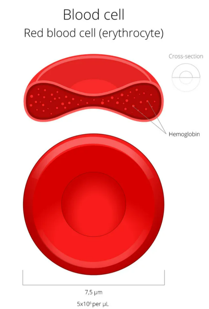
- Appearance in photomicrographs/diagrams:
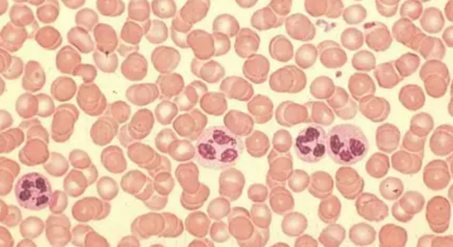
- Round, biconcave discs (doughnut-like shape).
- No nucleus (clear centre).
- Appear many in number, tightly packed.
- Stain light red/pink in microscope images.
- Key ID trick: Uniform size, pale centre, huge numbers.
⚪ White Blood Cells (WBCs / Leucocytes)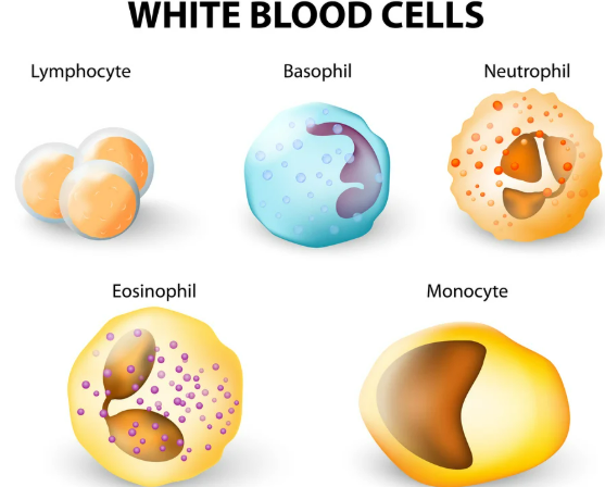
- Appearance in photomicrographs/diagrams:
- Much fewer than RBCs.
- Have a large, stained nucleus (dark purple/blue under stain).
- Irregular in shape (some round, some lobed).
- Larger in size compared to RBCs.
- Key ID trick: Look for darkly stained nucleus and bigger size.
📝 Summary Table
| Feature | RBC (Erythrocyte) | WBC (Leucocyte) |
|---|---|---|
| Shape | Biconcave disc | Irregular/round |
| Nucleus | Absent | Present (large, stained) |
| Size | Smaller | Larger |
| Number | Very many | Few |
| Staining | Light pink/red | Dark purple nucleus |
⚡ Quick Recap
RBC = small, many, no nucleus, pale centre.
WBC = large, few, dark nucleus.
👉 Mnemonic: “Red = Regular, White = With Nucleus”
Identifying Lymphocytes & Phagocytes
📌 Introduction
Both are types of white blood cells, but they look quite different under the microscope. Examiners often test whether you can tell them apart.
Lymphocytes
- Size → about same size as a red blood cell.
- Nucleus → very large, round, takes up most of the cell (thin rim of cytoplasm).
- Cytoplasm → narrow layer, appears as a thin blue “halo” around the nucleus.
- Function reminder → produce antibodies for specific defence.
👉 Tip: If the nucleus looks huge and dominates the cell → think “L” for Lymphocyte.
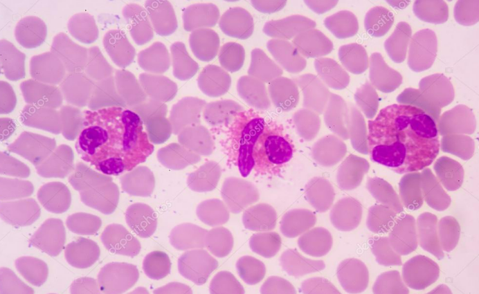
Phagocytes (e.g. neutrophils, macrophages)
- Size → much larger than red blood cells.
- Nucleus → irregular, lobed or kidney-shaped (not smooth and round).
- Cytoplasm → more abundant compared to lymphocytes.
- Function reminder → engulf and digest pathogens by phagocytosis.
👉 Tip: If the nucleus looks wriggly/lobed and there’s plenty of cytoplasm → it’s a Phagocyte.
📝 Comparison Table
| Feature | Lymphocyte | Phagocyte |
|---|---|---|
| Size | Same as RBC | Much larger than RBC |
| Nucleus | Large, round, fills most of cell | Lobed / irregular |
| Cytoplasm | Thin rim only | Large amount |
| Function | Antibody production | Phagocytosis |
⚡ Quick Recap
Lymphocyte → Little cell, Large nucleus
Phagocyte → Plenty of cytoplasm, Funny-shaped nucleus
👉 Mnemonic: “L = Little, P = Plenty”