CIE iGCSE Biology- 11.1 Gas exchange in humans- Study Notes- New Syllabus
CIE iGCSE Biology-11.1 Gas exchange in humans- Study Notes – New syllabus
CIE iGCSE Biology-11.1 Gas exchange in humans- Study Notes -CIE iGCSE Biology – per latest Syllabus.
Key Concepts:
Core
- Describe the features of gas exchange surfaces in humans, limited to: large surface area, thin surface, good blood supply and good ventilation with air
- Identify in diagrams and images the following parts of the breathing system: lungs, diaphragm, ribs, intercostal muscles, larynx, trachea, bronchi, bronchioles, alveoli and associated capillaries
- Investigate the differences in composition between inspired and expired air using limewater as a test for carbon dioxide
- Describe the differences in composition between inspired and expired air, limited to: oxygen, carbon dioxide and water vapour
- Investigate and describe the effects of physical activity on the rate and depth of breathing
Supplement
- Identify in diagrams and images the internal and external intercostal muscles
- State the function of cartilage in the trachea
- Explain the role of the ribs, the internal and external intercostal muscles and the diaphragm in producing volume and pressure changes in the thorax leading to the ventilation of the lungs
- Explain the differences in composition between inspired and expired air
- Explain the link between physical activity and the rate and depth of breathing in terms of: an increased carbon dioxide concentration in the blood, which is detected by the brain, leading to an increased rate and greater depth of breathing
- Explain the role of goblet cells, mucus and ciliated cells in protecting the breathing system from pathogens and particles
Features of Gas Exchange Surfaces in Humans
Gas exchange in humans happens in the alveoli, the tiny air sacs in the lungs. These structures are specially adapted to efficiently exchange gases like oxygen and carbon dioxide between the lungs and the bloodstream.
To do this well, gas exchange surfaces need four key features:
1. Large Surface Area
- The lungs contain millions of alveoli, each shaped like a tiny balloon.
- Together, these provide a huge surface area (around 70 m² in adults).
- This allows more oxygen to enter the blood and more carbon dioxide to leave.
2. Thin Surface (Short Diffusion Distance)
- Each alveolus has very thin walls – just one cell thick.
- Capillaries around alveoli are also thin.
- This ensures a short diffusion path, speeding up gas exchange.
3. Good Blood Supply
- Alveoli are surrounded by capillaries carrying deoxygenated blood.
- Oxygen diffuses into the blood, and carbon dioxide diffuses out.
- A steady blood flow keeps a steep concentration gradient.
4. Good Ventilation with Air
- Breathing in brings fresh oxygen-rich air, while breathing out removes CO₂-rich air.
- This keeps oxygen levels high and carbon dioxide levels low in the alveoli – supporting fast diffusion.
📌 Summary Table
Feature | Purpose |
---|---|
Large surface area | Increases space for gas exchange |
Thin surface | Allows quick diffusion of gases |
Good blood supply | Maintains steep concentration gradients |
Good ventilation | Keeps oxygen high and carbon dioxide low |
Parts of the Human Breathing System
This system helps us breathe in oxygen and breathe out carbon dioxide.
Let’s understand the major parts of the breathing system and what they do:

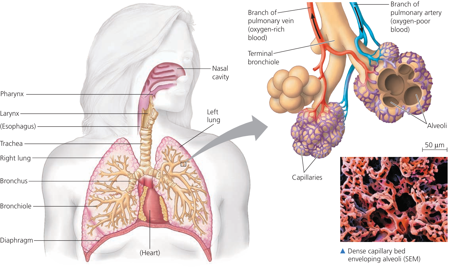
1. Lungs: Two large, spongy organs in the chest. Main site of gas exchange oxygen enters the blood and carbon dioxide leaves. Right lung is slightly larger.
2. Diaphragm: Dome-shaped muscle beneath the lungs. Moves down during inhalation to help lungs expand and moves up during exhalation to push air out.
3. Ribs: Protective cage around lungs and heart. Move up and out during inhalation, increasing chest volume.
4. Intercostal Muscles: Found between the ribs.
- External: Lift ribs during inhalation
- Internal: Help in forced exhalation
5. Larynx (Voice Box): Top of the trachea. Produces sound and prevents food from entering the windpipe.
6. Trachea (Windpipe): Connects larynx to bronchi. Contains cartilage rings to keep it open. Carries air to and from lungs.
7. Bronchi: Two tubes branching from trachea, one to each lung. Carry air deeper into lungs.
8. Bronchioles: Smaller tubes branching from bronchi. Spread air throughout lungs.
9. Alveoli: Tiny air sacs at ends of bronchioles. Site of gas exchange oxygen into blood, carbon dioxide out.
10. Capillaries (around Alveoli): Tiny blood vessels around alveoli. Carry blood in and out. Walls are one cell thick to allow fast exchange.
📌 Quick Recap Chart
Structure | Function |
---|---|
Lungs | Main organs for gas exchange |
Diaphragm | Contracts and relaxes to move air in and out |
Ribs | Protect lungs and assist breathing |
Intercostal muscles | Move ribs during inhalation/exhalation |
Larynx | Produces sound, routes air to trachea |
Trachea | Airway from throat to bronchi |
Bronchi | Carry air to each lung |
Bronchioles | Distribute air within lungs |
Alveoli | Site of gas exchange |
Capillaries | Transport gases to/from alveoli |
Investigating the Difference Between Inspired and Expired Air
🔍 Objective (Using Limewater to Test for Carbon Dioxide)
To compare the amount of carbon dioxide in inhaled (inspired) and exhaled (expired) air using a simple experiment with limewater.
Materials Needed
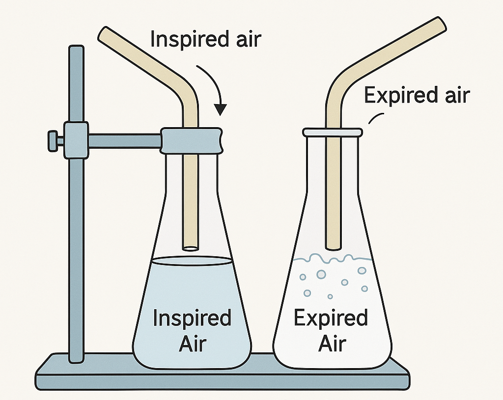
- Two test tubes or boiling tubes
- Fresh limewater (clear calcium hydroxide solution)
- Two straws or rubber tubes
- Clamp stand or test-tube rack
🧪 What is Limewater?
Limewater is a chemical test for carbon dioxide (CO₂). When CO₂ is bubbled through it, the solution turns milky or cloudy white due to the formation of calcium carbonate.
🧬 Experiment Setup
- Label two test tubes: one for inspired air, one for expired air.
- Fill both tubes with equal amounts of limewater.
Tube 1 (Inspired Air): Use a straw to bubble air from the surroundings (not from your mouth) through the limewater. This represents the air we inhale.
Tube 2 (Expired Air): Blow your exhaled breath through the limewater using a straw. This shows the air we exhale after respiration.
Observations
- Tube 1 (Inspired Air): Limewater remains clear or turns slightly cloudy → low CO₂
- Tube 2 (Expired Air): Limewater turns milky much faster → high CO₂
Conclusion
Expired air contains more carbon dioxide than inspired air. This proves that the body adds CO₂ during respiration, which is then removed when we exhale.
📌 Summary Table
Feature | Inspired Air | Expired Air |
---|---|---|
Carbon Dioxide Level | Low | High |
Effect on Limewater | Stays clear or slightly cloudy | Turns milky quickly |
💡 Scientific Link
This experiment visualizes gas exchange. Oxygen is taken in from inspired air, and carbon dioxide (a waste product of respiration) is added to expired air – which is exactly how the lungs function.
Differences in Composition Between Inspired and Expired Air
🔄 What Happens During Breathing?
When we inhale (inspire) air, we take in gases from the atmosphere. When we exhale (expire), the air has changed inside our lungs due to gas exchange.
📊 Key Differences in Gases
Gas | Inspired Air (Inhaled) | Expired Air (Exhaled) |
---|---|---|
Oxygen (O₂) | ~21% | ~16% |
Carbon Dioxide (CO₂) | ~0.04% | ~4% |
Water Vapour | Low (varies with humidity) | High (always moist) |
🧪 Explanation
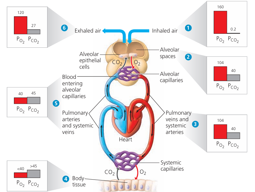
- Oxygen (O₂): Decreases in expired air because it is used by body cells during aerobic respiration.
- Carbon Dioxide (CO₂): Increases in expired air as it’s a waste product of respiration, released by cells and transported to the lungs for removal.
- Water Vapour: Always higher in expired air because air is humidified as it passes through moist surfaces in the lungs and airways.
🧠 Quick Summary
- Inspired air has more oxygen, less carbon dioxide, and is less humid.
- Expired air has less oxygen, more carbon dioxide, and is more humid.
Effects of Physical Activity on Breathing
🔬 What Happens During Physical Activity?
During exercise (e.g. running, cycling), your muscles need more oxygen and produce more carbon dioxide due to increased aerobic respiration. To meet this demand, your breathing system adjusts quickly.
Two Main Changes in Breathing
Feature | At Rest | During Exercise |
---|---|---|
Breathing Rate | 12–18 breaths/min | Up to 40–60 breaths/min |
Breathing Depth | Shallow | Deeper |
📈 Why Does This Happen?
Increased breathing rate = more breaths per minute
→ Brings in more oxygen and removes more CO₂
Increased breathing depth = more air per breath
→ Enables better gas exchange in alveoli
This ensures:
- Oxygen reaches muscles faster
- CO₂ is removed efficiently to prevent acidity
Trained athletes often have lower resting breathing rates but much higher efficiency during intense activity.
🧪 How to Investigate This
- Measure resting breathing rate (count breaths for 1 minute)
- Perform light exercise (e.g. jog on the spot for 2-3 min)
- Measure breathing rate again
- Observe both rate and depth increase
You may also notice faster chest movement, mild breathlessness, and recovery within minutes.
🧠 Key Idea
Physical activity increases both the rate and depth of breathing to deliver more oxygen and remove carbon dioxide faster — supporting the demands of active muscles.
Identifying Internal and External Intercostal Muscles

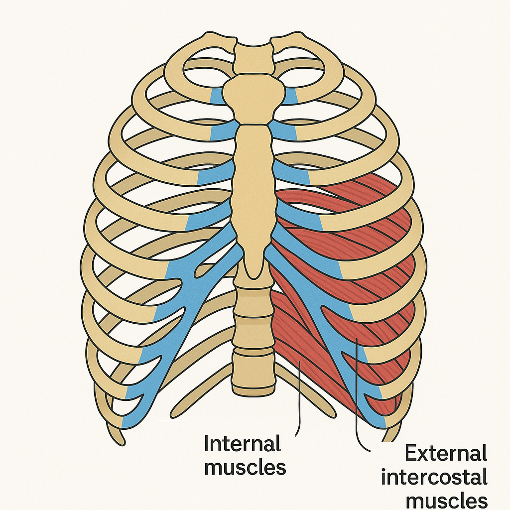
✅ What are Intercostal Muscles?
Intercostal muscles are the muscles found between the ribs.
They play a key role in breathing by helping move the ribcage during inhalation and exhalation.
🔄 Two Types:
Muscle Type | Location | Function |
---|---|---|
External Intercostal Muscles | Outside the ribcage (more superficial) | Pull ribs up and out during inhalation |
Internal Intercostal Muscles | Inside the ribcage (more deep) | Pull ribs down and in during forced exhalation |
Function of Cartilage in the Trachea
The trachea (windpipe) contains C-shaped rings of cartilage along its length.
✅ Main Function
Cartilage rings keep the trachea open and prevent it from collapsing – especially during breathing when air pressure fluctuates.
🔍 Why Is This Important?
When you inhale, air pressure inside the trachea drops. Without structural support, the tracheal walls could collapse inward, blocking airflow.
The C-shape of the cartilage rings allows the back of the trachea to stay soft and flexible enabling the oesophagus (located just behind the trachea) to expand when swallowing food.
Cartilage provides rigid support to the airway while still allowing flexibility for nearby organs like the oesophagus.
How Breathing Works: Ventilation of the Lungs
Breathing (also called ventilation) involves two main processes:
- Inhalation (breathing in)
- Exhalation (breathing out)
These depend on volume and pressure changes in the thorax, aided by the ribs, intercostal muscles, and diaphragm.
Inhalation (Inspiration)
Structure | Action | Effect |
---|---|---|
External intercostal muscles | Contract | Pull ribs up and out |
Internal intercostal muscles | Relax | No effect (in normal breathing) |
Diaphragm | Contracts and flattens | Increases thoracic volume |
Overall Effect: Thorax volume ↑ → Pressure inside thorax ↓ → Air flows into the lungs
Exhalation (Expiration)
Structure | Action | Effect |
---|---|---|
External intercostal muscles | Relax | Ribs move down and in |
Internal intercostal muscles | Contract (during forceful exhalation) | Pull ribs inward |
Diaphragm | Relaxes and domes upward | Decreases thoracic volume |
Overall Effect: Thorax volume ↓ → Pressure inside thorax ↑ → Air is pushed out
Air always moves from high pressure to low pressure. By changing the volume of the thorax, we change the pressure inside it — this is how air is drawn in and pushed out during breathing.
Differences Between Inspired and Expired Air
1. Oxygen (O₂)
Inspired air: ~21%
Expired air: ~16%
Some oxygen is absorbed into the bloodstream during gas exchange in the lungs for cellular respiration.
2. Carbon Dioxide (CO₂)
Inspired air: ~0.04%
Expired air: ~4%
Carbon dioxide is a waste product of respiration and is released from the blood into the alveoli to be exhaled.
3. Water Vapour (H₂O)
Inspired air: Varies (depends on humidity)
Expired air: Always high
Moist surfaces in the lungs cause air to become saturated with water vapour during exhalation.
4. Nitrogen (N₂)
Inspired air: ~78%
Expired air: ~78%
Nitrogen is inert and not used in respiration, so its proportion remains roughly unchanged.
5. Temperature
Inspired air: Varies (depends on environment)
Expired air: Warmer (close to body temperature)
Air warms up as it passes through the respiratory tract.
Summary Table
Component | Inspired Air (%) | Expired Air (%) | Notes |
---|---|---|---|
Oxygen (O₂) | ~21 | ~16 | Used in respiration |
Carbon Dioxide (CO₂) | ~0.04 | ~4 | Waste product of respiration |
Water Vapour | Variable | High | Increases due to moist lungs |
Nitrogen (N₂) | ~78 | ~78 | Unchanged, not involved in exchange |
Temperature | Cooler | Warmer | Air warms in respiratory system |
Physical Activity and Breathing Rate: The Role of Carbon Dioxide
What happens during physical activity?
- Muscles work harder and use more oxygen for aerobic respiration.
- They also produce more carbon dioxide (CO₂) as a waste product.
Effect on Blood CO₂ Levels
- Increased muscle activity = More CO₂ enters the bloodstream.
- This causes a rise in CO₂ concentration in the blood.
How the Brain Detects CO₂
- The medulla oblongata (in the brain) has chemoreceptors.
- These receptors detect the increased CO₂ levels in the blood.
How Breathing Responds
- The brain sends signals to the respiratory muscles:
- Diaphragm
- Intercostal muscles
- This leads to:
- Increased rate of breathing (more breaths per minute)
- Greater depth of breathing (deeper inhalations)
Purpose of This Response
- Removes excess CO₂ from the blood (via exhalation)
- Brings in more oxygen to meet the muscles’ increased demand
- Helps maintain homeostasis (stable internal environment)
Summary Flow
Protection of the Breathing System: Goblet Cells, Mucus & Ciliated Cells
1. Goblet Cells
Found in the lining of the airways (trachea and bronchi).
Their main function is to produce mucus.
- Role: Secrete sticky mucus that traps:
- Dust particles
- Pathogens (bacteria, viruses, fungi)
- Other harmful airborne particles
2. Mucus
A sticky fluid made mainly of water, glycoproteins, and salts.
Covers the surface of the respiratory epithelium.
- Role: Traps microbes and dust, preventing them from reaching the lungs.
- Acts as a first line of defence against infection and irritation.
3. Ciliated Cells
These are epithelial cells with tiny hair-like projections called cilia.
Located next to goblet cells in the lining of the airways.
- Role: The cilia beat rhythmically, moving the mucus (with trapped particles) upwards toward the throat.
- The mucus is then:
- Swallowed (where stomach acid kills pathogens)
- Or coughed/sneezed out
Combined Defence Mechanism
Component | Function |
---|---|
Goblet cells | Produce mucus to trap particles |
Mucus | Sticky barrier for dust/pathogens |
Ciliated cells | Sweep mucus out of airways |
Goblet cells make mucus → Mucus traps dust/pathogens → Cilia sweep mucus away → Pathogens removed before reaching the lungs