Home / iGCSE / iGCSE Biology 0610 / Summary Notes / CIE iGCSE Biology-14.1 Coordination and response- Study Notes

CIE iGCSE Biology-14.1 Coordination and response- Study Notes

CIE iGCSE Biology-14.1 Coordination and response- Study Notes- New Syllabus

CIE iGCSE Biology-14.1 Coordination and response- Study Notes – New syllabus

CIE iGCSE Biology-14.1 Coordination and response- Study Notes -CIE iGCSE Biology – per latest Syllabus.

Key Concepts:

Core

  • State that electrical impulses travel along neurones
  • Describe the mammalian nervous system in terms of:
     (a) the central nervous system (CNS) consisting of the brain and the spinal cord
     (b) the peripheral nervous system (PNS) consisting of the nerves outside of the brain and spinal cord
  • Describe the role of the nervous system as coordination and regulation of body functions
  • Identify in diagrams and images sensory, relay and motor neurones
  • Describe a simple reflex arc in terms of: receptor, sensory neurone, relay neurone, motor neurone and effector
  • Describe a reflex action as a means of automatically and rapidly integrating and coordinating stimuli with the responses of effectors (muscles and glands)
  • Describe a synapse as a junction between two neurones

Supplement

  • Describe the structure of a synapse, including the presence of vesicles containing neurotransmitter molecules, the synaptic gap and receptor proteins
  • Describe the events at a synapse as:
     (a) an impulse stimulates the release of neurotransmitter molecules from vesicles into the synaptic gap
     (b) the neurotransmitter molecules diffuse across the gap
     (c) neurotransmitter molecules bind with receptor proteins on the next neurone
     (d) an impulse is then stimulated in the next neurone
  • State that synapses ensure that impulses travel in one direction only

CIE iGCSE Biology-Concise Summary Notes- All Topics

Electrical Impulses and Neurones

Key Statement:

Electrical impulses travel along neurones to transmit information rapidly around the body.

🔍 What Are Electrical Impulses?

  • Tiny electrical signals generated by neurones (nerve cells).
  • Used by the nervous system to send messages quickly and precisely.

🧠 Neurones – The Signal Carriers

FeatureDescription
NeuronesSpecialised cells that carry nerve impulses.
AxonThe long part of a neurone where the impulse travels.
DirectionImpulses travel in one direction: from dendrites → cell body → axon terminals.

⚙️ Function of Electrical Impulses

  • Allow quick communication between:
    • Receptors (e.g. in skin)
    • Spinal cord/brain (processing centre)
    • Effectors (muscles or glands)
  • Enable rapid responses to changes in the environment (stimuli).
📝Note:
Neurones = nerve cells
Impulses = electrical messages
Travel fast to coordinate actions
Allow reactions like pulling away from hot objects

The Mammalian Nervous System

Overview:

  • The nervous system is the body’s fast communication system.
  • It detects stimuli, processes information, and coordinates responses through electrical impulses.

(a) Central Nervous System (CNS)

ComponentFunction
BrainActs as the control centre, processes information, stores memories, and makes decisions.
Spinal CordConnects the brain to the rest of the body and coordinates reflex actions.

🧠 The CNS:

    • Is protected by the skull (brain) and vertebrae (spinal cord).
    • Processes all incoming sensory information and sends out responses via motor neurones.


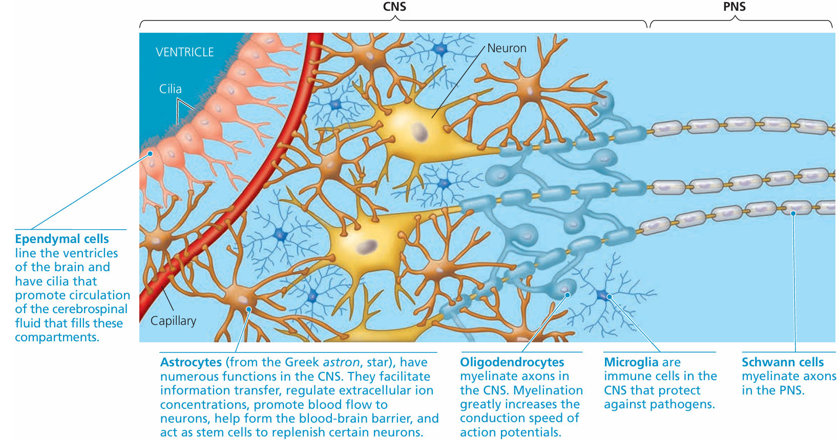
(b) Peripheral Nervous System (PNS)

ComponentFunction
Nerves outside the CNSCarry impulses to and from the brain and spinal cord.
Includes:
– Sensory neurones: carry messages to the CNS
– Motor neurones: carry commands from the CNS to effectors

🟢 The PNS:

  • Connects the CNS to the limbs and organs.
  • Allows the body to respond quickly to stimuli.
  • Plays a role in both voluntary and involuntary responses.

📘 Summary:

SystemIncludesMain Role
CNSBrain + Spinal cordProcesses and coordinates responses
PNSAll nerves outside CNSSends messages to and from the CNS
🗒️Note:
CNS = Brain + Spinal cord (control centre)
PNS = Nerves (communication network)
Together, they detect, process, and respond to stimuli

Role of the Nervous System: Coordination and Regulation

Key Function:

The nervous system is responsible for the coordination and regulation of body functions by detecting changes (stimuli), processing information, and producing appropriate responses.

🔍 What is Coordination?

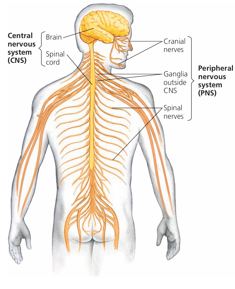
Coordination means making sure that all body parts work together smoothly.
The nervous system ensures that different organs and systems respond in a controlled and balanced way.

🧠 Example:

If you touch something hot, your hand quickly pulls away – muscles, skin receptors, spinal cord, and brain all work together instantly.

⚙️ What is Regulation?

Regulation means keeping internal body conditions stable and functioning properly.
The nervous system helps control:

  • Heart rate
  • Breathing rate
  • Movement
  • Reflexes
  • Voluntary and involuntary actions

How the Nervous System Works:

StepAction
1Stimulus (e.g., pain, temperature) is detected by receptors.
2Electrical impulses are sent through sensory neurones to the CNS.
3Brain or spinal cord processes the information.
4A response is sent through motor neurones to effectors (muscles or glands).
5The effector produces the appropriate response (e.g., muscle contraction, hormone release).

Types of Neurones & How to Identify Them

1. Sensory Neurone

Function: Carries electrical impulses from receptors (e.g., skin) to the central nervous system (CNS)
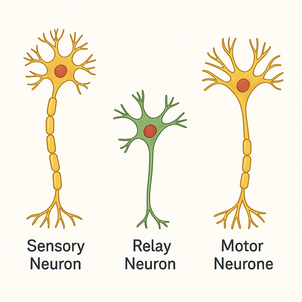
FeatureHow to Identify
Cell bodyLocated off to the side (in a swelling called the dorsal root ganglion)
DirectionFrom receptor → CNS
StructureLong dendron (from receptor), short axon (to CNS)

2. Relay Neurone (Interneurone)

Function: Carries impulses within the CNS, usually between sensory and motor neurones

FeatureHow to Identify
Cell bodyIn the middle of the neurone
DirectionFrom sensory neurone → motor neurone
StructureShort axons and dendrites, found only in brain and spinal cord

3. Motor Neurone

Function: Carries impulses from the CNS to effectors (muscles or glands)

FeatureHow to Identify
Cell bodyLocated at one end (within the spinal cord)
DirectionFrom CNS → effector
StructureLong axon, short dendrites
Neurone TypeDirection of ImpulseCell Body LocationConnected To
SensoryReceptor → CNSSide (ganglion)Receptor to CNS
RelayCNS → CNSCentreBetween sensory & motor
MotorCNS → EffectorStart of neuroneCNS to muscle/gland

Simple Reflex Arc

Definition:

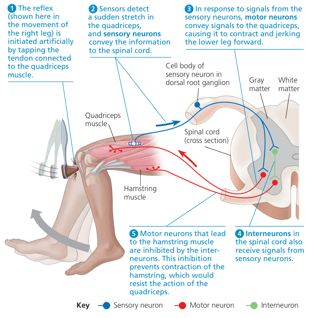
A reflex arc is the pathway taken by a nerve impulse during a reflex action — a quick, automatic response to a stimulus that protects the body from harm.

🔄 Steps in a Simple Reflex Arc:

StepDescription
1. StimulusA change in the environment is detected (e.g., touching a hot object).
2. ReceptorA sensory receptor in the skin detects the stimulus (e.g., heat or pain).
3. Sensory NeuroneCarries the impulse from the receptor to the spinal cord.
4. Relay NeuroneLocated in the spinal cord (CNS), passes the impulse from sensory to motor neurone.
5. Motor NeuroneCarries the impulse from the spinal cord to the effector.
6. EffectorA muscle or gland that carries out the response (e.g., the muscle contracts to pull your hand away).

🧠 Key Feature of Reflexes:

  • Very fast (no brain involvement in the decision).
  • Helps prevent injury by producing immediate protective actions.
📌Note:
Stimulus → Receptor → Sensory neurone → Relay neurone → Motor neurone → Effector
Reflex actions are involuntary and bypass the brain for faster response.

Reflex Action – Definition and Description

What is a Reflex Action?

A reflex action is an automatic and rapid response to a stimulus, designed to protect the body from harm.

🔄 Key Features:

FeatureDescription
AutomaticHappens without conscious thought (involuntary).
RapidInvolves minimal nerve pathway – no delay from brain processing.
ProtectiveHelps avoid injury or harm (e.g., blinking, pulling hand away from heat).
CoordinationConnects stimulus with the appropriate effector quickly.

🧠 How It Works:

  • Receptors detect the stimulus (e.g. pain).
  • Sensory neurones carry the impulse to the spinal cord.
  • Relay neurones in the spinal cord transfer the signal.
  • Motor neurones carry the impulse to the effectors (muscles/glands).
  • Effectors carry out the response (e.g. muscle contracts, gland secretes).

🧷 Example:

Touching a hot object
→ Skin receptors detect heat
→ Sensory neurone sends impulse to spinal cord
→ Relay neurone passes message
→ Motor neurone sends impulse to biceps
→ Muscle contracts → hand pulls away

🗒️ Note:
Reflex = Fast + Involuntary + Protective
Stimulus → Response via spinal cord
Effectors = Muscles or glands

What is a Synapse?

Definition:

A synapse is a tiny gap or junction between two neurones, where nerve impulses are transmitted chemically from one neurone to the next.

🧠 Key Points:

FeatureDescription
StructureA synapse is the gap between the axon terminal of one neurone and the dendrite of the next.
TransmissionThe impulse is passed across the gap using chemical messengers called neurotransmitters.
DirectionImpulses only travel in one direction across a synapse.

🔄 How It Works:

  • An electrical impulse reaches the end of the first neurone (axon terminal).
  • This triggers the release of neurotransmitters into the synaptic gap.
  • The neurotransmitters diffuse across the gap.
  • They bind to receptors on the second neurone.
  • A new electrical impulse is generated in the second neurone.
🗒️Note:
Synapse = Gap between neurones
Neurotransmitters = Chemicals that cross the gap
Direction = Always one-way

Structure of a Synapse

Definition:

A synapse is a tiny gap between two neurones where nerve impulses are transmitted chemically using neurotransmitters.

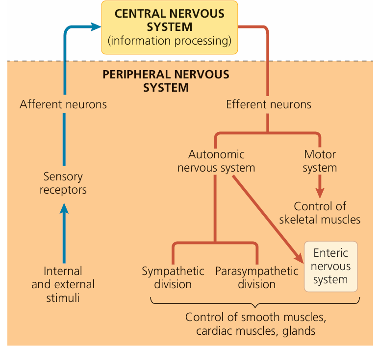
🔍 Key Components of a Synapse:

PartDescription
Presynaptic neuroneThe neurone sending the signal. Its axon terminal contains vesicles filled with neurotransmitters.
Synaptic vesiclesSmall sacs that store and release neurotransmitter molecules (e.g. acetylcholine).
Synaptic cleftThe tiny gap (20–40 nm) between the presynaptic and postsynaptic neurones where neurotransmitters diffuse.
Postsynaptic neuroneThe receiving neurone with receptor proteins that bind neurotransmitters.
Receptor proteinsLocated on the postsynaptic membrane; they detect neurotransmitters and trigger a new impulse.

🔄 Transmission Process:

  • Electrical impulse arrives at presynaptic terminal.
  • Vesicles release neurotransmitters into the synaptic cleft.
  • Neurotransmitters diffuse across the gap.
  • They bind to receptor proteins on the postsynaptic neurone.
  • This stimulates a new electrical impulse.
🧷 Note:
Synapse = Presynaptic neurone + Synaptic cleft + Postsynaptic neurone
Vesicles = Store neurotransmitters
Receptors = Detect neurotransmitters and trigger response

Events at a Synapse

What is a Synapse?

A synapse is the junction between two neurones, where signals are passed from one neurone to the next using chemical messengers called neurotransmitters.

🔄 Step-by-Step Events at a Synapse:

StepDescription
(a) Impulse arrival and neurotransmitter releaseWhen an electrical impulse reaches the axon terminal of the first (presynaptic) neurone, it triggers vesicles to move to the membrane and release neurotransmitter molecules into the synaptic gap.
(b) Diffusion across the synaptic gapThe neurotransmitter molecules then diffuse across the synaptic cleft (the small gap between the two neurones).
(c) Binding to receptor proteinsThe neurotransmitters bind to specific receptor proteins on the membrane of the postsynaptic neurone.
(d) New impulse generatedThis binding stimulates a new electrical impulse in the next neurone, allowing the signal to continue.

📌 Summary:

  • Transmission across a synapse is chemical (neurotransmitters)
  • Direction is one-way: from presynaptic → postsynaptic neurone
  • Synapses slow down impulses slightly but allow flexibility in response

One-Way Transmission at Synapses

Key Statement:

Synapses ensure that impulses travel in one direction only.

Explanation:

A synapse is the gap between two neurones, where information is passed using chemical messengers called neurotransmitters.

  • Only the presynaptic neurone has vesicles that store and release neurotransmitters.
  • Only the postsynaptic neurone has receptor proteins to detect and respond to neurotransmitters.

Because of this arrangement, impulses can only pass from the presynaptic → postsynaptic neurone, and not in reverse.

📘 Summary Table:

StructureFound inRole
Neurotransmitter vesiclesPresynaptic neuroneReleases signal
Receptor proteinsPostsynaptic neuroneDetects and responds to signal
Note:
Synapses are built for one-way flow
Vesicles → neurotransmitters → receptors
This ensures signals only move forward
Scroll to Top