Identification of Main Blood Vessels in Diagrams
(a) Main Blood Vessels to and from the Heart
| Vessel | Function | Direction of Blood Flow |
|---|---|---|
| Vena Cava | Brings deoxygenated blood from the body to the right atrium | Into the heart |
| Pulmonary Artery | Carries deoxygenated blood from the right ventricle to the lungs | Away from the heart |
| Pulmonary Vein | Brings oxygenated blood from the lungs to the left atrium | Into the heart |
| Aorta | Carries oxygenated blood from the left ventricle to the body | Away from the heart |
📌 In Diagram:
• The vena cava connects to the right atrium (usually shown top right side).
• The pulmonary artery exits the right ventricle and leads to the lungs.
• The pulmonary vein enters the left atrium from the lungs.
• The aorta exits from the left ventricle and arches to the body.
(b) Main Blood Vessels to and from the Lungs
| Vessel | Function | Direction of Blood Flow |
|---|---|---|
| Pulmonary Artery | Carries deoxygenated blood from the heart to the lungs for gas exchange | From heart → lungs |
| Pulmonary Vein | Carries oxygenated blood from the lungs to the heart | From lungs → heart |
📌 In Diagram:
• Pulmonary arteries branch from the heart to both lungs.
• Pulmonary veins return to the left atrium from each lung.
(c) Main Blood Vessels to and from the Kidneys
| Vessel | Function | Direction of Blood Flow |
|---|---|---|
| Renal Artery | Supplies oxygenated blood with waste materials to the kidney | From heart → kidney |
| Renal Vein | Carries filtered, deoxygenated blood from the kidney back to the heart | Kidney → heart (via vena cava) |
📌 In Diagram:
• The renal artery enters each kidney (thicker wall, high pressure).
• The renal vein leaves each kidney (thinner wall, lower pressure).
🧾 Summary Table
| Organ | Blood Vessels | Type of Blood |
|---|---|---|
| Heart | Vena Cava, Aorta, Pulmonary Artery & Vein | Deoxygenated & Oxygenated |
| Lungs | Pulmonary Artery & Vein | Deoxygenated → Oxygenated |
| Kidney | Renal Artery & Renal Vein | Oxygenated & Filtered |
How the Structure of Arteries and Veins is Related to Blood Pressure

Arteries – High Pressure
Function: Carry oxygenated blood (except pulmonary artery) away from the heart under high pressure.
🔬 Structural Adaptations:
- Thick muscular walls: Withstand high pressure from heartbeats.
- Elastic tissue: Allows stretching and recoiling to maintain pressure.
- Small lumen: Maintains pressure and fast flow.
- No valves: Not needed due to strong forward pressure.
📌 Why?
Blood enters arteries forcefully, so they must be strong and elastic to handle pressure surges and keep blood moving efficiently.
Veins – Low Pressure
Function: Return deoxygenated blood (except pulmonary vein) back to the heart under low pressure.
🔬 Structural Adaptations:
- Thinner walls: No need to withstand high pressure.
- Less elastic tissue: No stretching/recoiling needed.
- Large lumen: Reduces resistance and allows easier flow.
- Valves present: Prevent backflow, especially in limbs.
📌 Why?
Because blood moves slowly in veins, valves and skeletal muscle contractions are needed to keep it flowing in the right direction.
🧾 Summary Table
| Feature | Arteries | Veins |
|---|---|---|
| Wall Thickness | Thick, muscular | Thin, less muscular |
| Elastic Tissue | Present | Little or absent |
| Lumen Size | Narrow | Wide |
| Valves | Absent | Present |
| Blood Pressure | High | Low |
| Direction of Flow | Away from heart | Towards heart |
How the Structure of Capillaries is Related to Their Functions
Capillaries are the smallest blood vessels in the body and are crucial for the exchange of substances between the blood and surrounding tissues.
🧪 Main Functions of Capillaries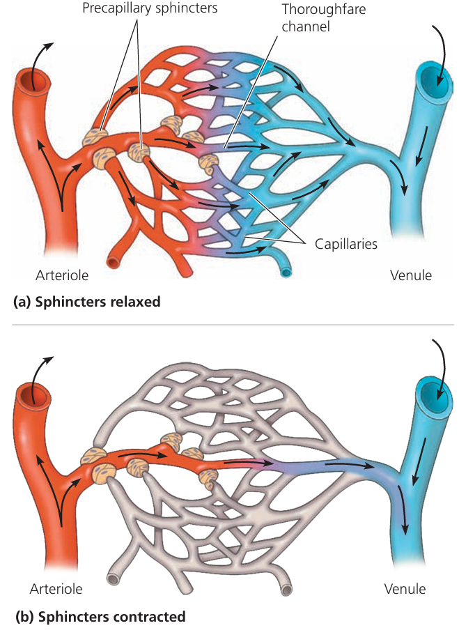
- Exchange of oxygen and carbon dioxide between blood and body cells
- Delivery of nutrients like glucose and amino acids to cells
- Removal of waste products such as urea and CO₂
- Absorption of nutrients in the small intestine
- Exchange of gases in alveoli of the lungs
🧬 Structural Features & Their Functions
| Structural Feature | How it Supports Capillary Function |
|---|---|
| Very thin walls (one cell thick) | Allows for rapid diffusion of gases and nutrients. |
| Single layer of endothelial cells | Minimizes diffusion distance for efficient exchange. |
| Extensive branching network | Increases surface area and access to all cells. |
| Extremely narrow diameter | Forces red blood cells to pass in single file for better gas exchange. |
| Slow blood flow | Allows time for exchange of materials. |
| Permeable walls | Allow plasma to leak out and form tissue fluid. |
| Located between arterioles and venules | Main site of exchange in circulation. |
📌 Why Capillary Structure Is Ideal for Exchange
Capillaries are perfectly adapted for their role. Their tiny size, thin walls, and wide distribution allow them to:
- Deliver oxygen and nutrients efficiently
- Remove CO₂ and waste products
- Allow hormones to reach target tissues
Main Blood Vessels to and from the Liver
The liver is one of the most important organs in the human body. It acts like a biochemical factory processing nutrients, detoxifying chemicals, and producing bile. To perform these functions, the liver receives blood from two main sources and sends blood out through one main vein.
1. Hepatic Artery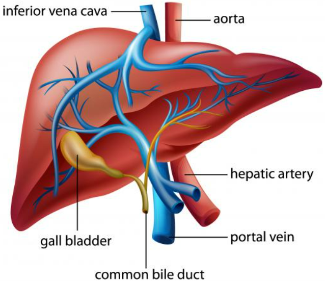
- Function: Brings oxygen-rich blood from the heart to the liver.
- Source: Originates from the aorta via the celiac artery.
- Purpose: Supplies liver cells (hepatocytes) with oxygen for cellular respiration and energy production.
2. Hepatic Portal Vein
- Function: Brings nutrient-rich blood from the digestive organs (like stomach and intestines) to the liver.
- Source: Formed by veins from the stomach, intestines, spleen, and pancreas.
- Purpose: Allows the liver to:
- Process absorbed glucose, amino acids, and fatty acids
- Store excess nutrients
- Detoxify harmful substances like alcohol or drugs
3. Hepatic Vein
- Function: Carries deoxygenated, processed blood from the liver to the heart.
- Destination: Drains into the inferior vena cava.
- Purpose: Removes blood that has already been filtered and processed by the liver.
🧠 Summary Table
| Blood Vessel | Type of Blood | Direction | Key Function |
|---|---|---|---|
| Hepatic artery | Oxygenated | Heart → Liver | Supplies liver cells with oxygen |
| Hepatic portal vein | Nutrient-rich, low oxygen | Intestines → Liver | Delivers nutrients and toxins for processing |
| Hepatic vein | Deoxygenated, processed | Liver → Heart (via vena cava) | Drains blood from liver |