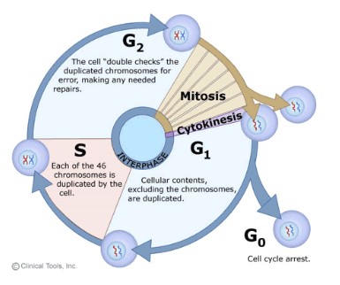
Topic 1 : Cells – 1.6 Cells division

Topic 1 Cell
1.6 Cell division

Cell cycle

  • G1 phase: increase cytoplasm volume, organelle production and protein synthesis (normal growth)
  • S phase: DNA replication
  • G2 phase: increase cytoplasm volume, double the amount of organelle and protein synthesis (prepare for cell division)
  • M phase: Mitosis
  • Interphase: consists of the parts of the cell cycle that don’t involve cell division (G1,S and G2 phase)
  • G0 phase: resting phase where the cell leaves the cell cycle and has stopped dividing. Cell carries out all normal functions without the need of dividing. e.g. brain cell
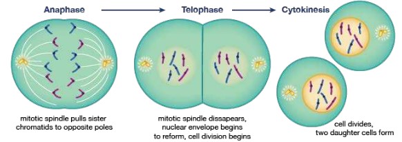
Prophase:

  • DNA supercoil: chromatin condenses and becomes sister chromatids, which are visible under the light microscope.
  • Nuclear membrane is broken down and disappeared.
  • Centrosomes move to the opposite poles of the cell and spindle fibers begin to form.

Metaphase:

  • Spindle fibers (microtubules) from each of the centrosomes attach to the centromere of sister chromatids.
  • Chromatids line up in the equator.

Anaphase:

  • Contraction of the spindle fibers cause the separation of the sister chromatids.
  • The chromatids are now considered as chromosomes.
  • Chromosomes move to opposite poles of the cell

Telophase:

  • Chromosomes uncoil to become chromatin.
  • Spindle fibers break down and new nuclear membrane reform at opposite pole.

Cytokinesis in animal cells:

  • a cleavage furrow forms when the plasma membrane is pulled inwards around the equator by the contractile proteins actin and myosin
  • Once the invagination reaches the centre the membrane pinches off and to new cells are formed

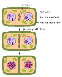
Cytokinesis in plant cells:

  • In plant cells tubular structures are formed by vesicles along the equator of the cell
  • This continues until two layers of membrane exist across the equator, which develop into the plasma membrane of the two new cells
  • Vesicles bring pectin and other substances and deposit these between the two membranes through exocytosis forming the cell plate
  • Cellulose is then brought and deposited by exocytosis between the membranes as well, forming the new cell walls
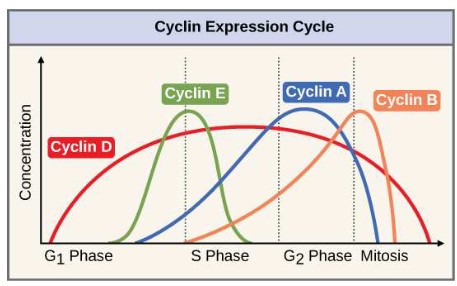
Cyclins:

  • Cyclins are a family of proteins that control the progression of cells through the cell cycle.
  • It is used to mark the checkpoints between two stages
  • Cells cannot progress to the next stage of the cell cycle unless the specific cyclin reaches its threshold
  • Cyclin binds to enzyme called cyclin-dependent kinases
  • This enzyme then trigger the signal to move on to the next stage.

Mutagens and oncogenes:

  • Tumors (cancer) are the result of uncontrolled cell division, which can occur in any organ or tissue.
  • These abnormal growths can either be localized (primary tumours), meaning they do not move to other part of your body. These tumours are benign.
  • If the cancer cells detach and move elsewhere into the body (secondary tumours), they are called malignant and are more life threatening
  • Diseases due to malignant tumours are known as cancer
  • Metastasis is the movement from a primary tumour to set up secondary tumours in other parts of the body
  • Cancer is usually caused by genetic abnormalities due to a variety of different sources called carcinogens or due to inheritance or errors in DNA replication.
  • Carcinogens are agents that can cause cancer, such as viruses, X-Rays, UV Radiation and many chemical agents
  • Mutagens are agents that can cause mutations in one’s DNA which can lead to cancer
  • In cancer two types of genes are usually affected, oncogenes and tumor suppressor genes.
  • Oncogenes are genes that control cell cycle and cell division.
  • Tumor suppressor genes usually control replication and the cell cycle. In cancer cells these genes are generally inactivated causing a loss of normal function.
Scroll to Top