Question
The electron micrographs show a typical prokaryote and a mitochondrion.
Prokaryote Mitochondrion
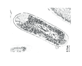
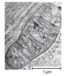
Compare and contrast the structure of a typical prokaryotic cell with that of a mitochondrion. [4]
Explain how mitochondria could have been formed from free living prokaryotes. [2]
▶️Answer/Explanation
Ans:
a
differences
a.prokaryote has cell wall but mitochondrion does not ✔
b.mitochondrion has double membrane whereas prokaryote has single membrane
OR
«Gram negative» bacteria have cell wall between two membranes whereasmitochondria has intermembrane space between two membranes ✔
c.mitochondrion has cristae/invaginations of inner membrane but prokaryote does not
OR
prokaryote «may have» flagella/pili/«slime» capsule which mitochondria do not have ✔
similarities
d.70S ribosomes in both ✔
e.DNA in both / loop of DNA in both / naked DNA in both ✔
f.shape similar/both rod shaped/OWTTE
OR
size of both is similar/both about 3 μm long ✔
g.both are membrane-bound/OWTTE ✔
b
a.endocytosis/engulfing of prokaryote by a larger/another/anaerobic prokaryote/cell ✔
b.double membrane of the mitochondrion is the result of endocytosis
OR
inner membrane of mitochondrion from engulfed cell and outer from food vacuole ✔
c.«engulfed prokaryotic cell» was aerobic/respired aerobically/consumed oxygen
OR
«engulfed prokaryotic cell» provided energy/ATP ✔
d.«engulfed prokaryotic cell» not destroyed/not digested
OR
«endo»symbiotic/mutualistic relationship developed ✔
e.«engulfed prokaryotic cell» had its own DNA/own «70S» ribosomes ✔
Question
Draw the ultrastructure of a prokaryotic cell based on electron micrographs. [3]
▶️Answer/Explanation
Ans
a.cell wall;
b.plasma membrane; Clearly shown as a separate line under the cell wall or theinner line
c.cytoplasm AND 70S ribosomes; Do not allow (small) circles
d.nucleoid/naked DNA;
e.plasmid
OR
pili
OR
flagella/flagellum;

Question
The diagram shows some of the structures in an animal cell.
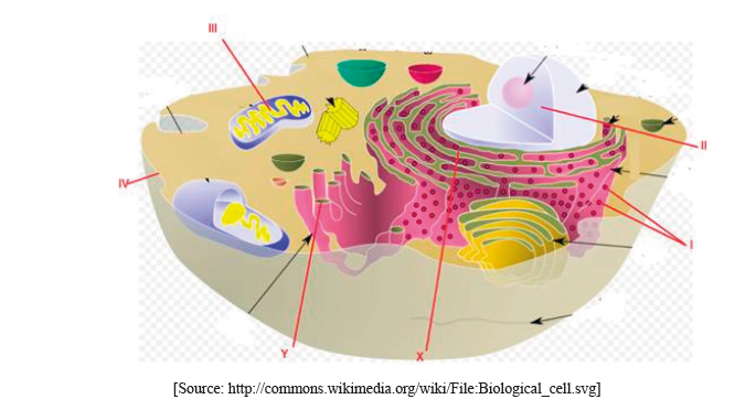
(i) Label structures I, II, III and IV.
I.
II.
III.
IV.
(ii) State one function of structure III.
Explain how materials are transported within a cell between structures X and Y.
▶️Answer/Explanation
(i) Award [1] for any two of the following correctly labeled.
I. ribosomes
II. nucleus (do not accept nuclear membrane)
III. mitochondrion
IV. plasma/cell membrane
(ii) ATP production/site of aerobic respiration (do not accept energy production)
(protein) material transported by vesicles;
from rER to Golgi apparatus/complex/body/membrane;
vesicles bud off from rER/fuse with Golgi membrane (due to membrane fluidity);
Do not accept vacuole(s).