Topic 11.3: The Kidney and Osmoregulation
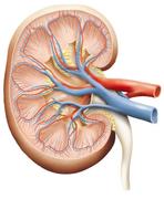 image from National Institute of Diabete
In the Kidneys and Osmoregulation unit we will learn how the kidney’s maintain homeostasis, the ability of the body to maintain a stable internal environment despite a changing environment. We will learn how the kidneys maintain the volume of body fluids., maintain the balance of salt ions in body fluids and excrete harmful nitrogen-containing molecules, such as urea, ammonia, and uric acid. We will also learn what happens when the kidney does not function properly.
This unit will last 4 school days.
Essential idea:
- All animals excrete nitrogenous waste products and some animals also balance water and solute concentrations.
Nature of science:
- Curiosity about particular phenomena—investigations were carried out to determine how desert animals prevent water loss in their wastes. (1.5)
11.3 U 1 Animals are either osmoregulators or osmoconformers.
- Define osmoregulator and osmoconformer.
- List three example osmoregulator animals and three example osmoconformer animals.
Osmolarity refers to the solute concentration of a solution
Osmoregulators
- maintain a constant internal solute concentration, even if they live in a marine environment with a very different osmolarity
- All terrestrial animals, freshwater animals and some marine organisms are osmoregulators because they maintain constant internal solute concentration, even when living in marine environments with very different osmolarities
- Typically these organisms maintain their solute concentration at about one third of the concentration of seawater and about 10 times that of fresh water
Osmoconformers
- animals that have similar internal solute concentration in comparison to the solute concentration of their surrounding environment
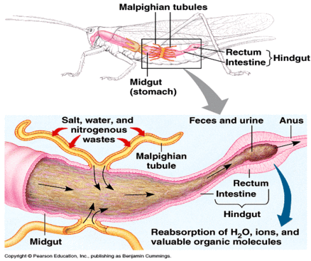
11.3 U 2 The Malpighian tubule system in insects and the kidney carry out osmoregulation and removal of nitrogenous wastes.
- Define osmoregulation.
- State the nitrogenous waste products found in insects and mammals.
- Outline the structure and function of the Malpighian tubule system.
Osmoregulation is a form of homeostasis whereby the concentration of hemolymph, or blood in the case of animals with closed circulatory systems, is kept within a certain range. When animals break down amino acids, the nitrogenous waste product is toxic and needs to be excreted.
Arthropods have circulating fluid, known as hemolymph, that combines the characteristics of tissue fluid and blood. In insects, the waste product is usually in the form of uric acid and in mammals it is in the form of urea. Insects have tubes that branch off from their intestinal tract. These are known as Malpighian tubules.
Cells lining the tubules actively transport ions and uric acid from the hemolymph into the lumen of the tubules. This draws water by osmosis from the hemolymph through the walls of the tubules into the lumen. The tubules empty their contents into the gut. In the hindgut most of the water and salts are reabsorbed while the nitrogenous waste is excreted with the feces
11.3 U 3 The composition of blood in the renal artery is different from that in the renal vein.
- State the functions of the kidney.
- Distinguish between osmoregulation and excretion.
- List 4 substances that are found in higher concentration in the renal artery than in the renal vein.
- Compare the relative glucose, oxygen and carbon dioxide concentrations between the renal artery and the renal vein.
- State that plasma proteins are not filtered by the kidney so should be present in the same concentration in the renal artery and renal vein.
Both arteries and veins (in the renal system or anywhere else) are types of blood vessels. Arteries carry blood away from the heart; veins return blood to the heart.
- In the renal artery blood enters the kidney
- In the renal vein blood leaves the kidney
Substances that are present in HIGHER amounts in the renal artery than the renal vein include:
- toxins and other substances that are ingested and absorbed but are not fully metabolised by the body e.g. betalain pigments in beets and also drugs
- excretory waste products including nitrogenous waste products, mainly urea
Other things removed from the blood by the kidney that are not excretory products include:
- excess water, produced by cell respiration or absorbed from food in the gut
- excess salt, absorbed from food in the gut
NOTE: the kidney is the filter of the body – thus the substances that need to be removed from the body are present in higher amounts in the blood vessel entering the kidney (the renal artery).
The metabolic activity of the kidney itself also causes a difference between the composition of blood in the renal artery and the renal vein. Blood leaving the kidney through the renal vein is deoxygenated relative to the renal artery because kidney metabolism requires oxygen. It also has a higher partial pressure of CO2 because this is a waste product of metabolism. Even though glucose is normally filtered and then entirely reabsorbed, some glucose is used by the metabolism of the kidney and therefore the concentration is slightly lower in the renal vein compared to the renal artery
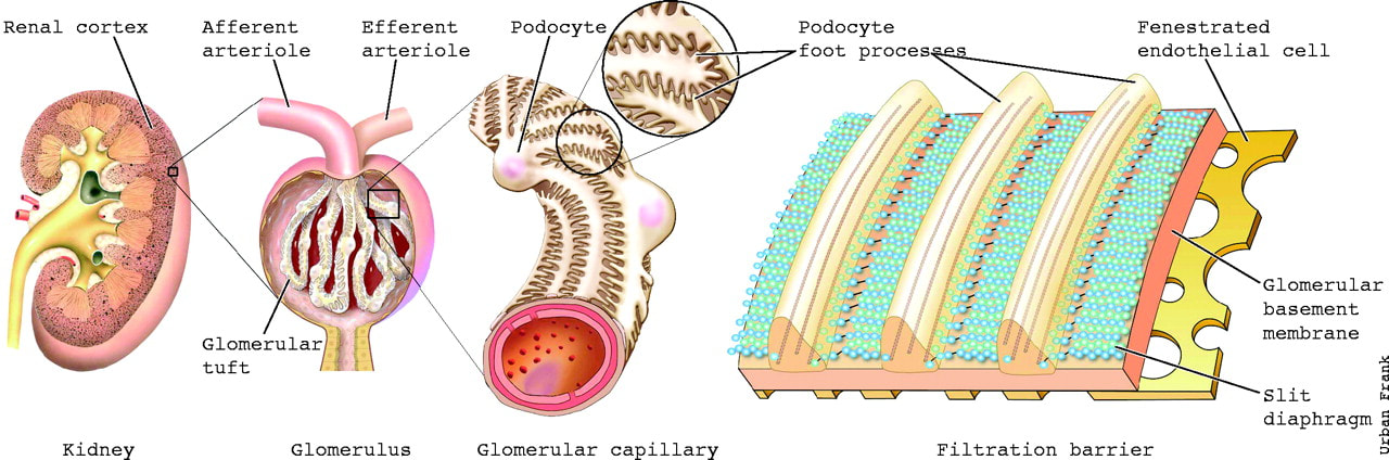
11.3 U 4 The ultrastructure of the glomerulus and Bowman’s capsule facilitate ultrafiltration.
- Outline the cause and effect of high blood pressure in the kidney glomerulus.
- List solutes found in glomerular filtrate.
- Define filtrate and ultrafiltration.
- Explain why plasma proteins and blood cells are not part of glomerular filtrate.
- On a glomerulus diagram, label the basement membrane, fenestrations, podocyte foot processes, podocytes.
- Outline the role of fenestration, the basement membrane and podocytes in ultrafiltration.
- Describe the relationship between the glomerulus and Bowman’s capsule.
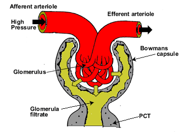
Ultrafiltration is the non-specific filtration of the blood as it enters the Bowman’s capsule of the kidney in which hydrostatic pressure created by the high pressure in the glomerulus (capillaries) forces a liquid against a semi-permeable membrane.
- blood in capillaries is at high pressure in many of the tissues of the body, and the pressure forces some of the plasma out through the capillary wall, to form tissue fluid
- the pressure in the capillaries of the glomerulus are particularly high and the capillary wall is particularly permeable, so the volume forced out is about 100 times greater than in other tissues
- the fluid forced out is called glomerular filtrate
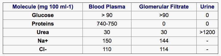
- the concentration of Na+ ions, Cl- ions, glucose and urea all remain about the same in both the composition of blood plasma and the filtrate, however protein (mg) differs enormously
- this separation of particles differing in size by a few nanometers is called ultrafiltration
- all particles with a relative molecular mass below 65,000 atomic mass units pass through
- the permeability to larger molecules depends on their shape and charge
- almost all proteins are retained in the blood, along with all blood cells
The glomerulus:
- the basement membrane separates the capillaries
- there are gaps in the wall of the capillaries called fenestrations
- smaller projections from the membrane are podocyte food processes, which attach the podocytes (specialised epithelial cells) to the membrane
- the podocytes function as a barrier through which waste products are filtered from the blood
There are three main parts to the ultrafiltration system:
- Fenestrations between the cells in the wall of the capillaries – these are about 100nm in diameter and they allow fluid to escape, but not blood cells
- The basement membrane that covers and supports the wall of the capillaries – it is made of negatively-charged glycoproteins, which form a mesh – it prevents plasma proteins from being filtered out, due to their size and negative charges
- Podocytes forming the inner wall of the Bowman’s capsule – these cells have extensions that wrap around the capillaries of the glomerulus and many short side branches called foot processes – very narrow gaps between the foot processes help prevent small molecules from being filtered out of the blood in the glomerulus
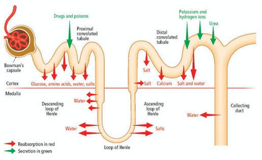
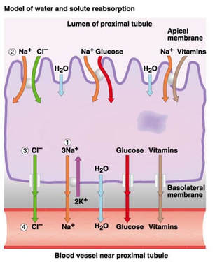
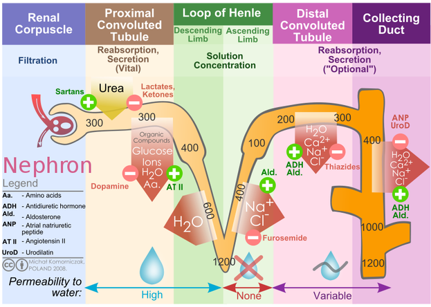
11.3 U 5 The proximal convoluted tubule selectively reabsorbs useful substances by active transport.
- List substances in the glomerular filtrate that are reabsorbed in the proximal convoluted tubule.
- Explain why cells lining the lumen of the proximal convoluted tubule have microvilli and many mitochondria.
- Outline the mechanism of selective reabsorption of sodium ions, chloride ions, glucose and water.
While ultrafiltration is non-specific with regards to the molecules that leave the blood (based on size, no specific channels), selective reabsorption in the proximal convoluted tubule (PCT) is very specific with regards to the molecules that are reabsorbed.
- the glomerular filtrate flows into the proximal convoluted tubule
- the volume of glomerular filtrate produced per day is huge – about 180dm-3
- most reabsorption of glucose and other substances mainly occurs in the first part of the nephron – the proximal convoluted tubule
Methods use to reabsorb substances in the proximal convoluted tubule include:
Sodium ions:
- moved by active transport from filtrate to space outside the tubule
- they then pass to the peritubular capillaries
- pump proteins are located in outer membrane of tubule cells
Chloride ions:
- attracted from filtrate to space outside the tubule because of charge gradient set up by active transport of sodium ions
Glucose:
- co-transported out of filtrate and into fluid outside the tubule, by co-transporter proteins in outer membranes of tubules cells
- sodium ions move down concentration gradient from outside tubule into tubule cells
- this provides energy for glucose to move at the same time to fluid outside the tubule
- the same process is used to reabsorb amino acids
By the end of the proximal tubule all glucose and amino acids and 80% of the water, sodium and other mineral ions have been absorbed.
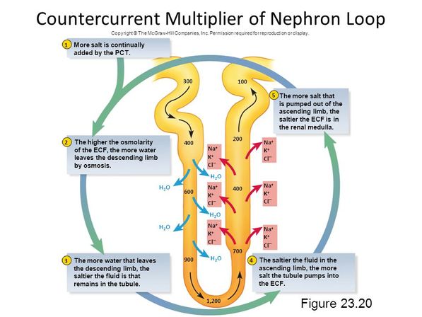
11.3 U 6 The loop of Henle maintains hypertonic conditions in the medulla.
- State the overall function of the loop of Henle.
- Outline the role of interstitial fluid in osmoregulation.
- Describe the structure and function of the descending limb of the loop of Henle.
- Describe the structure and function of the ascending limb of the loop of Henle.
- Describe why the loop of Henle is a countercurrent multiplier system.
The role of the loop of Henle is to create a solute concentration gradient in the medulla of the kidney.
- The descending loop of Henle is permeable to water but impermeable to salt ions.
- The ascending loop of Henle is permeable to salt ions but impermeable water.
- As the filtrate flows up the ascending loop Na+ ions are pumped out of the filtrate into the interstitial fluid of the medulla thus increasing the solute concentration in the medulla.
- As the filtrate flows down the descending loop (before the ascending loop)water flows out of the filtrate into the interstitial fluid of the medulla by osmosis following the concentration gradient creating by pumping Na+ ions out of the ascending loop.
- Therefore the medulla has a very high concentration of Na+ ions; maximum level in humans is 1200 mOsm (milliosmole).
- This system is countercurrent because the flow of the filtrate in the descending and ascending loop is in opposite directions. This allows for a greater concentration gradient to be created in the medulla.
- The filtrate now leaves the loop of Henle and enters the collecting duct where the water level is fine tuned.
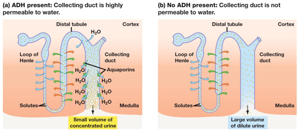
11.3 U 7 ADH controls reabsorption of water in the collecting duct.
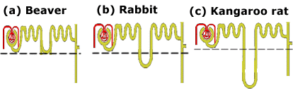
- Outline the relationship between habitat and length of the loop of Henle.
- Outline the relationship between habitat and relative medullary thickness.
11.3 U 8 The length of the loop of Henle is positively correlated with the need for water conservation in animals
- Outline the tonicity of filtrate entering the distal convoluted tubule from the loop of Henle.
- Outline the of low blood solute concentration on the volume of urine produced, solute concentration in the urine, permeability of the distal convoluted tubule and collecting duct to water and volume of water reabsorbed.
- Outline the of high blood solute concentration on the volume of urine produced, solute concentration in the urine, permeability of the distal convoluted tubule and collecting duct to water and volume of water reabsorbed.
- Outline the source and function of ADH in osmoregulation.
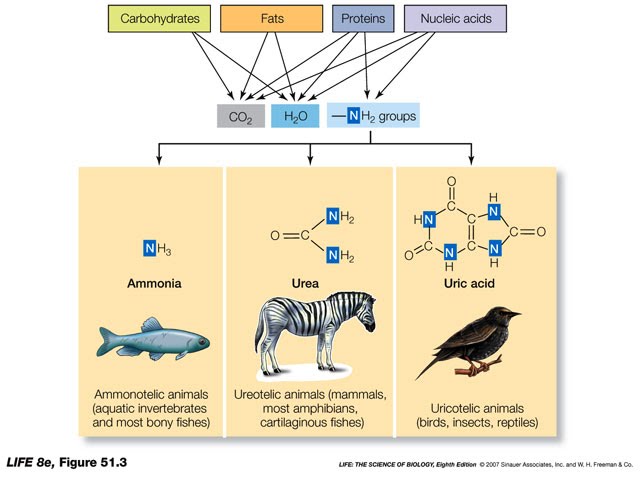
11.3 U 9 The type of nitrogenous waste in animals is correlated with evolutionary history and habitat.
- Outline the production and effect of ammonia in animals.
- State the nitrogenous waste products released by: aquatic organisms, terrestrial organisms, marine mammals, amphibians, birds and insects.
- Compare urea and uric acid.
When animals breakdown amino and nucleic acids, nitrogenous waste is formed in the form of ammonia. Ammonia is highly basic, toxic and can be very reactive. Marine and freshwater organisms can release the ammonia directly into the surrounding water.
- Terrestrial organisms convert ammonia into a less toxic form (urea or uric acid) before excretion. What form depends on the animal’s evolutionary history and habitat.
- Amphibians release waste as ammonia as larvae and urea as adults
- Birds and insects release ammonia as uric acid. Uric acid does not require water and is highly concentrated. This is beneficial to these organisms as they do not have to carry the extra water around (less energy for flight)
- Mammals release their waste in the less toxic form known as urea
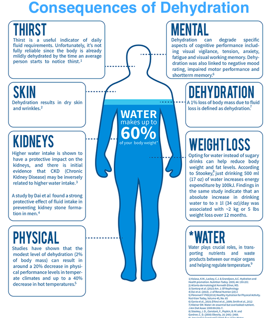
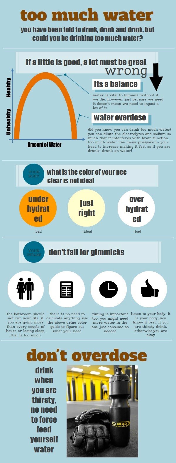
11.3 A 1 Consequences of dehydration and overhydration.
- Outline the causes and consequences of dehydration.
- Outline the causes and consequences of overhydration
- Dehydration is the consequence of losing large amounts of water through excessive sweating, diarrhea, vomiting or urination, without the loss of equal amounts of solutes
- The body fluids become hypertonic
- This results in dark coloured urine, lethargy, a raised heart rate and low blood pressure
- Overhydration results when too much water is consumed and the body fluids become hypotonic
- This results in behavioural changes, confusion, drowsiness, delirium, blurred vision, muscle cramps, nausea, and in acute cases, seizures, coma and finally death
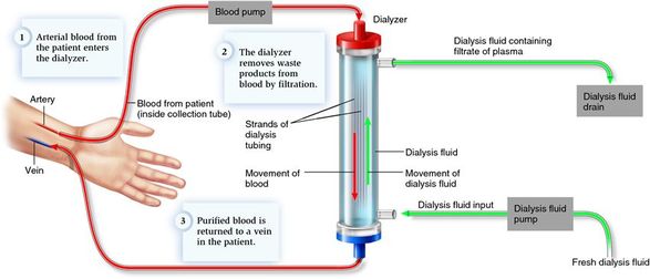
11.3 A 2 Treatment of kidney failure by hemodialysis or kidney transplant.
- List two common causes of kidney failure.
- Outline the process of hemodialysis.
- Outline the process of kidney transplant.
- Outline the treatment of kidney stones by ultrasound.
Hemodialysis
- Blood is drawn from a vein in the arm and is passed through a dialysis machine for 3 to 4 hours, 3 times per week
- As the blood flows through the machine, it passed next to a semi-permeable membrane that contains dialysis fluid
- Pores in the membrane allow small particles to diffuse in either direction; however, the pores are too small for plasma proteins and cells to pass through and therefore remain in the blood
- Dialysis fluid (dialysate) has the following characteristics that allows the blood to be cleaned
- No urea or waste – These products diffuse from the blood into the fluid
- Ideal concentrations of glucose and other metabolites – ideal concentrations of these molecules is therefore created in the blood of the patient through diffusion onto or out of the blood
- High concentration of calcium and low concentration of potassium – Excess potassium is extracted out of the blood and calcium is added to the blood
- HCO3- is added to the blood to make it more basic
- Correct total solute concentration – Allows for excess water to diffuse from the blood
Kidney Transplant
- A better long term treatment for a kidney that is not working properly is a kidney transplant
- Either a living person provides one of their kidneys for transplant or kidneys are removed from someone that has recently died in order to give two kidneys to two individuals
- It is essential to have matching blood types and tissue matches to minimize the risk of rejection
- The kidney is grafted to the lower abdomen and the renal artery renal vein and ureter are connected to the individual
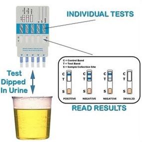
11.3 A 3 Blood cells, glucose, proteins and drugs are detected in urinary tests.
- Define urinalysis.
- Outline the use of a urine test strip in detection of diabetes, kidney damage and drug use.
- Outline the microscopic examination of urine for detection of infection, kidney stones or kidney tumors.
Urine tests are used to detect abnormalities and possible diseases
- Blood cells – blood cells in the urine is an indicator of a variety of diseases, including some cancers and infections
- Glucose – excess glucose in the urine is almost always an indicator of diabetes
- Proteins – Larger amounts of proteins in the urine are an indicator of kidney disease. Small amounts of some hormones such as insulin is normal
- Drugs – Many drugs pass from the body into the urine, therefore a urine test can be conducted to detect drug users in sports or for recreational users tested by the police
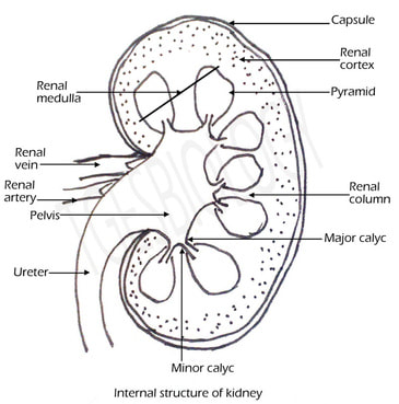
11.3 S 1 Drawing and labelling a diagram of the human kidney.
- Draw a diagram of a human kidney.
- Label the renal artery, renal vein, cortex, medulla, renal pelvis and ureter on a diagram of the human kidney.
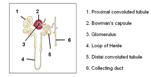
11.3 S 2 Annotation of diagrams of the nephron.
- Define nephron.
- Annotate a diagram of the nephron with the following structures and associated functions: Bowman’s capsule, proximal convoluted tubule, Loop of Henle,. distal convoluted tubule, collecting duct, afferent arteriole, glomerulus, efferent arteriole, peritubular capillaries, vasa recta and venules.
The nephron is the functional unit of the kidney, with each nephron being comprised of the following components:
- Bowman’s capsule
- Proximal convoluted tubule
- Loop of Henle
- Distal convoluted tubule