Topic 11.4: Sexual Reproduction

In the Sexual Reproduction unit we will explore gamete formation in both male and female humans. We will then learn how these gametes are fused together to form a zygote and eventually a fetus. We will also look at the hormones that are involved in fetal development.
This unit will last 4 school days
Essential idea:
- Sexual reproduction involves the development and fusion of haploid gametes.
Nature of science:
- Assessing risks and benefits associated with scientific research—the risks to human male fertility were not adequately assessed before steroids related to progesterone and estrogen were released into the environment as a result of the use of the female contraceptive pill. (4.8)
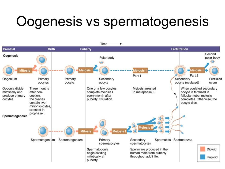
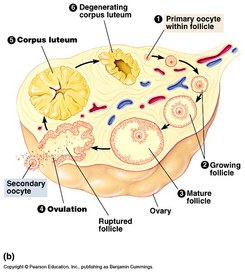
OOGENESIS:
- oogenesis is the production of egg cells in the ovaries
- oogenesis starts in the ovaries of a female fetus
- germ cells in the fetal ovary divide by mitosis and the cells formed move to distribute themselves through the cortex of the ovary
- when the fetus is four/five months old, these cells grow and start to divide by meiosis
- by the seventh month, they are still in the first division of meiosis and a single layer of cells, called follicle cells, has formed around them
- no further development takes place until after puberty
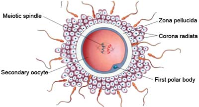
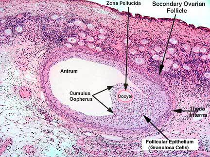
- the cell that has started to divide by meiosis, together with the surrounding follicle cells, is called a primary follicle
- there are about 400,000 in the ovaries at birth
- no more primary follicles are produced, but at the start of each menstrual cycle, a small batch are stimulated to develop by FSH
- usually one goes on to become a mature follicle, containing a secondary oocyte
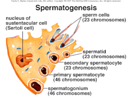
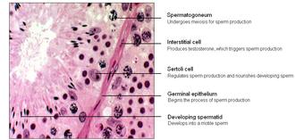
SPERMATOGENESIS:
- spermatogenesis is the production of sperm
- it happens in the testes, which are composed of a mass of narrow tubes, called seminiferous tubules, with small groups of cells filling the gaps between the tubules
- these gaps are called interstices, so the cells in them are interstitial cells
- they are sometimes called Leydig cells
- the seminiferous tubules are also made of cells
- the outer layer of cells is called the germinal epithelium, with the most mature stages closest to the fluid-filled centre of the seminiferous tubule
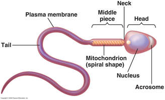
- cells that have developed tails are called spermatozoa
- in the wall of the tubule are large nurse cells, called Sertoli cells, which nourish the sperm cells
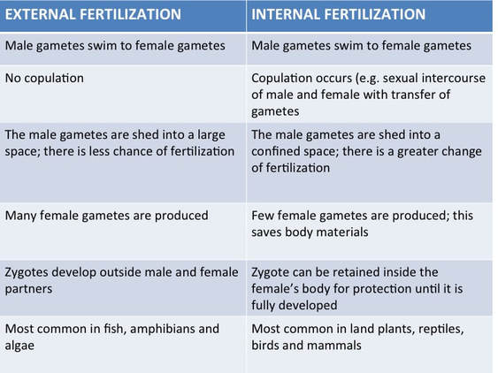
Internal Fertilization
The fertilization process occurs within the body of a female is called internal fertilization. It is a specialization for the protection of egg, but it depends on the birth method . Reptiles and birds have thick shell, covering the egg to protect it from dehydration and destruction. However, fertilization occurs inside the body otherwise, sperm has to enter through a thick wall.
Animals such as mammals also have internal fertilization, where embryo develops inside mother, which enhances the protection to the embryo1.
Internal fertilization facilitates the survival of the embryo, selective fertilization, and longer protection and minimizes the wastage of gametes.
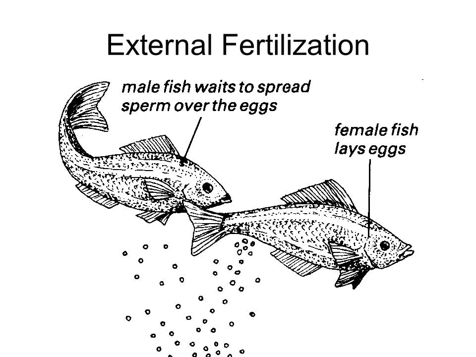
External Fertilization
In external fertilization fusion of sperm and egg occurs externally of the female body. External fertilization needs water to facilitate their fertilization, so it occurs in wet environments. Female and male gametes are released to the water, and male gamete is mostly mobile. This type of fertilization can be seen in lower plants. The advantage of external fertilization is that it produces a large number of offspring due to the external hazards. So survival of the embryo is comparatively lower. Amphibians and fish are examples for these types of animals.
The internal fertilization process occurs within the body of female whereas, in external fertilization, fusion of sperm and egg occurs externally of the female body.
• After the internal fertilization, egg will come out of the body having a thick shell whereas, in external fertilization, eggs are produced with thin tertiary membrane or without membrane .
• External fertilization needs water, whereas internal fertilization does not need water to fertilize.
• Organisms involved in external fertilization have mobile male gametes with flagella, whereas organisms involved in internal fertilization has immobile male gametes.
• In internal fertilization, wastage of gametes is lower, whereas wastage of gametes is higher in external fertilization.
• Organisms that involved in internal fertilization produce lower number of gametes, whereas organisms involved in external fertilization produce a large number of gametes.
• Survival of organisms that involved in internal fertilization is higher than the survival of organisms involved in external fertilization.
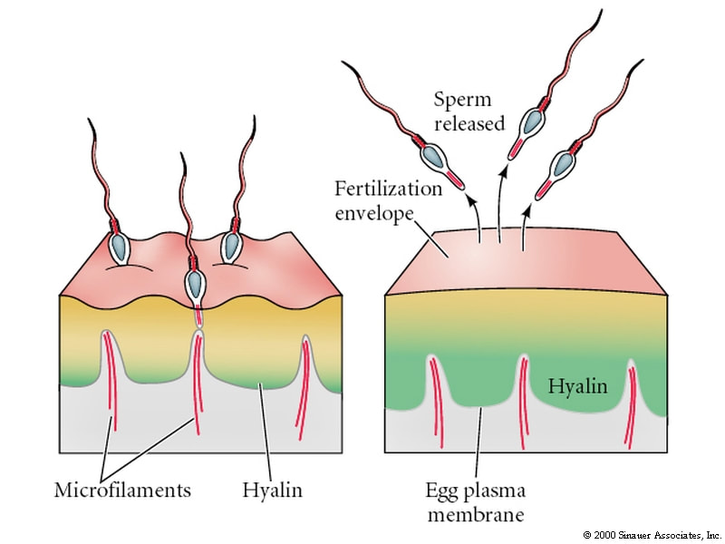
Acrosome Reaction
- sperm migrates through follicle cells (corona radiata)
- sperm binds to zona pellucida glycoproteins
- exocytosis of acrosomal hydrolytic enzymes
- enzymatic digestion through zona pellucida (proteins + polysaccharides)
Egg and sperm membrane fusion
- acrosomal reaction exposes sperm membrane
- binding proteins of egg and sperm match
- binding triggers egg membrane depolarization as Na+ gates open
- fast block to polyspermy
Cortical reaction
- depolarization triggers release of egg G proteins
- G proteins trigger release of Ca++ from ER
- Ca++ triggers fusion of cortical granules with egg membrane
- enzymes released by exocytosis catalyze hardening of zona pellucida
- slow block to polyspermy
Fertilization
- egg microvilli endocytotically take in sperm nucleus
- envelopes of both nuclei dissolve and share in common spindle apparatus
- first mitosis of embryo = cleavage division
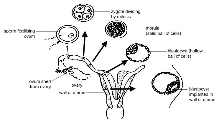
After the male and the female gametes combine to form a zygote, the zygote divides by mitosis to form a two-cell embryo. They two cells grow and replicate their DNA, and undergo another cell division through mitosis to form a four-cell embryo. As the embryo is developing, it is moving along the fallopian tube towards the uterus.
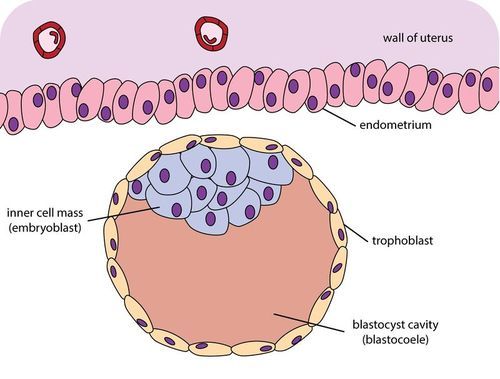
The four-cell embryo continues to divide by cell division until it reaches 16 to 32 cells; called the morula. After continued cell divisions a blastocyst consisting of 100 to 128 cells is formed and is ready for implantation into the endometrium.
The blastocyst consists of an inner cell mass that will develop into the body of the embryo, a group of cells surrounding the embryo called the trophoblast that will develop into the placenta, and a fluid-filled cavity called the blastocoel.. The outer layer of cells will develop finger-like projections that will allow the embryo to penetrate the uterine wall during implantation.
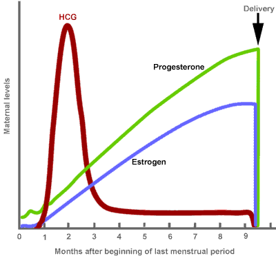
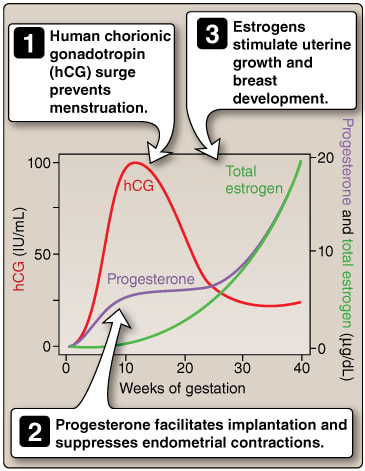
When a human embryo is implanted into the endometrium or the uterine lining, it starts to produce the hormone, human chorionic gonadotrophin (HCG).
embryo secretes HCG
- which acts like pituitary LH,maintaining corpus luteum
- which produces estrogen and progesterone, maintaining endometrium and cervical mucus/plug
- stimulates growth of placenta and uterine enlargement
- inhibits menstruation
by second trimester, HCG declines
- corpus luteum disintegrates
- placenta produces progesterone/estrogen
presence of HCG
- can be detected in mother’s urine by ELISA (enzyme-linked immunosorbent assay)
- as a positive test of pregnancy commonly used in pregnancy test kits
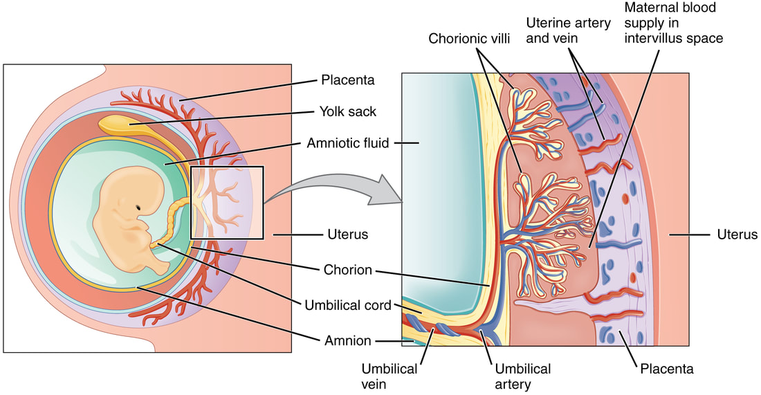
The placenta develops from the trophoblast layer of the blastocyst. When developed three blood vessels contained within umbilical cord connect the placenta to the growing fetus.
Two umbilical arteries carry deoxygenated blood and waste away from the fetus to the placenta. As maternal blood enters the placenta it leaves the arteries and enters the inter-villous space, where it pools and surrounds the placental villi. The placental villi are finger-like fetal tissues that have a large surface area for the exchange of materials such as gases, nutrients and wastes.
Fetal blood that circulates in capillaries within the villi and microvilli is very close to the surface, allowing for efficient exchange of materials between the fetal and maternal blood. Materials such as oxygen, nutrients and vitamins diffuse into the fetal capillaries from the maternal blood in the inter-villous space, while carbon dioxide and wastes diffuse out of the fetal capillaries into the inter-villous space. One umbilical vein carries oxygenated and nutrient rich blood back to the fetus from the placenta.
The cells that separate the fetal and maternal blood form a semi-permeable placental barrier. Materials are exchanged between the maternal and the fetal blood in the placenta.
Note: maternal and fetal blood is never mixed together.
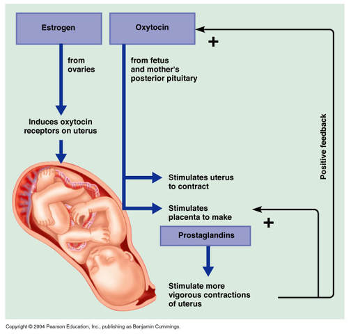
When the pregnancy is at term, the fetus secretes hormones that signal the placenta to stop producing progesterone (progesterone inhibits the secretion of oxytocin by the pituitary gland). Oxytocin secreted by the anterior pituitary gland stimulates the muscle fibers in the uterus to begin to contract. As the muscles in uterus contract, mechanoreceptors in the uterine wall signal the pituitary to produce more oxytocin. More oxytocin increases the frequency and intensity of the contractions, thus stimulating the production of even more oxytocin. This is an example of positive feedback.
Contractions of the muscles of the uterus will cause the amniotic sac to break, releasing the amniotic fluid (This is when the “water breaks” in childbirth). Relaxation of the muscles in the cervix causes it to dilate, eventually allowing the increasing contractions to push the baby out through the vagina and the cervix. he placenta is expelled “afterbirth” about 15 minutes after the baby is born.
labor:
- first stage of birth
- progesterone decrease/estrogen increase: triggers formation of oxytocin receptors in uterus
- prostaglandins: produced by fetus/placenta initiate uterine contractions
- positive feedback: uterine contractions, via nervous system, stimulate production of oxytocin, a hormone, by posterior pituitary into blood
- oxytocin: binds to uterine receptors, stimulating:
- uterine contractions
- prostaglandin production by placenta
delivery:
- second stage of birth
- uterine contractions force fetal head against cervix, which dilates
- fetal head move through cervix, into vagina, exits
afterbirth:
- third stage of birth: placenta expelled
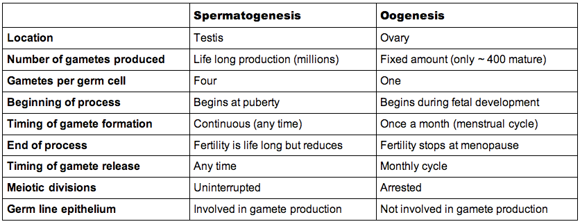
The process of gametogenesis occurs in the reproductive organs (gonads) of the male and female
- In males, the gametes are produced within the seminiferous tubules of the testes
- In females, the gametes are produced by the ovaries
The male and female reproductive gametes (sperm and egg) have specialised structures which reflect their functions
- The male gamete (sperm) is small and motile and only contributes the male’s haploid nucleus to the zygote
- The female gamete (egg) is large and non-motile and contributes all the organelles and cytoplasm to the zygote