Topic 6.1: digestion AND ABSORPTION
In the Digestion and Absorption unit you will learn how large food particles can not be absorbed through cell membranes that line the small intestine. These large molecules must be broken down into smaller molecules through the mechanical and chemical mechanisms.
This unit will last 3 school days.
Essential idea:
- The structure of the wall of the small intestine allows it to move, digest and absorb food.
Nature of science:
Use models as representations of the real world—dialysis tubing can be used to model absorption in the intestine. (1.10)
- Explain the use of models in physiology research.
- State two examples of model systems used to study digestion.
- State limitations of using model systems in physiology research.
6.1.U1 The contraction of circular and longitudinal muscle of the small intestine mixes the food with enzymes and moves it along the gut.
- Outline the role of peristalsis in the digestive process.
Peristalsis is the principal mechanism of movement in the oesophagus, although it also occurs in both the stomach and gut. Continuous segments of longitudinal smooth muscle rhythmically contract and relax. Food is moved unidirectionally along the alimentary canal in a caudal direction (mouth to anus)
- contraction of circular and longitudinal muscle of the small intestine helps mix (mechanical digestion) the food with enzymes and moves the semi-digested food (bolus) along the gut in a process called peristalsis
- These muscles are made up of smooth muscle
- The process by which continual waves of contraction and relaxation (peristalsis) of the circular and longitudinal muscle occur along the muscle layers surrounding the small intestine is controlled by the autonomic nervous system
- Food is transported slowly through the small intestine to allow for maximum digestion and absorption of nutrients
U 6.1.U2 The pancreas secretes enzymes into the lumen of the small intestine.
- List the name and substrate of the three major classes of enzymes secreted by the pancreas.
- Endopeptidases – example trypsin (breaks apart the peptide bond between amino acids in polypeptides)
- Lipases – catalyzes the hydrolysis of lipids (triglycerides and phospholipids)
- Amylases – digestion of starch.
- Pancreatic juice is alkaline (basic) to allow enzymes to work at an optimal pH (around 7-8 in the small intestine)
The pancreas is controlled by the enteric nervous system and through hormones produced and released by the stomach. Enzymes are produced by ribosomes in the pancreatic gland cells, excreted by exocytosis into smaller ducts, which converge to form the pancreatic duct. Pancreatic juice flows through the pancreatic duct into the lumen of the small intestine
6.1.U3 Enzymes digest most macromolecules in food into monomers in the small intestine.
- List the name, substrate and product of four pancreatic enzymes that hydrolyze food in the small intestine.
- List the name, substrate and product of six enzymes produced by gland cells in the small intestine wall.
- Describe why enzymes produced by gland cells in the small intestine wall often remain immobilized in the cell membrane.
Enzymes released by the small intestines break down macromolecules into smaller molecules called monomers through catabolic reactions (hydrolysis is the type of specific reaction).
The rate of reaction is actually slow due to the body temperature. Therefore enzymes increase the rate of breakdown by acting as biological catalysts. They work by hydrolysis of bonds to form monomers. Digestion is completed by enzymes located on microvilli and lumen of the small intestine.
The following is a table showing the enzyme, source, substrate, products
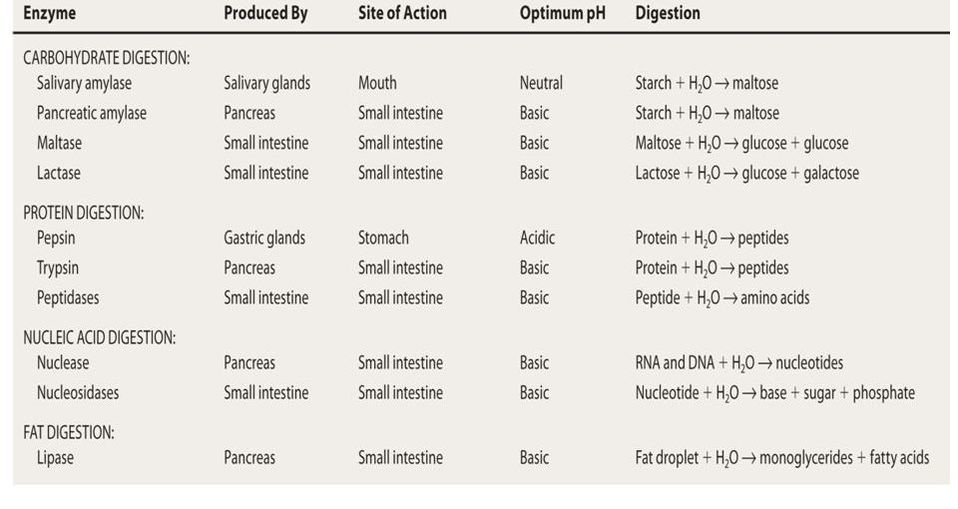
- Please note that some carbohydrates such as cellulose cannot be digested by humans as we lack the enzymes needed to break down cellulose
- This is why a number grass eating animals have specialized digestive systems to breakdown cellulose
- Cellulose is insoluble dietary fibre that helps move waste through our digestive system and prevents constipation
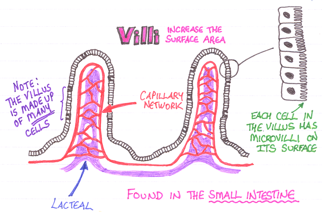
6.1.U4 Villi increase the surface area of epithelium over which absorption is carried out
- List three adaptations that increase the surface area for absorption on the small intestine.
- Draw the villi as viewed in cross section.
- Label the following on a diagram of a villi: capillary, epithelial cell, lacteal, and goblet cell.
- State the function of the following villi structures: capillary, epithelial cell, lacteal, and goblet cell.
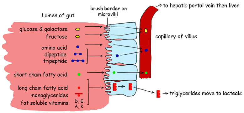
Absorption is the process by which the nutrients of food move from the small intestine into the bloodstream. It requires the digestion of food into monomers that are small enough to pass across cell membranes. The structure of the villus is related to its role in the absorption and the transport of the products of digestion.
- Villi are finger-like projections that make the surface of the small intestine look highly folded. These projections increase the surface area (by about 10X) available for absorption (the process of taking substances into the cells and blood).
- Microvilli are small hair-like projections attached to the villi to further increase surface area.
- The outermost layer of the villi is thin epithelial cells to allow nutrients to easily move across a short distance into the blood.
- A dense network of capillaries close to the epithelium allow a large amount of nutrients to move into the blood.
- Lacteals, which are a part of the lymphatic system, run up the middle of the villi. The lacteal allows for the absorption of the products of lipid digestion which are not easily absorbed by the capillaries.
- Protein channels and pumps are present in the membranes of epithelium to help with absorptio
 |  |
6.1.U5 Villi absorb monomers formed by digestion as well as mineral ions and vitamins. (Oxford Biology Course Companion page 283)
- Define absorption.
- List materials absorbed by the villi cells of the small intestine.
Absorption – process where small molecules and nutrients pass into the blood vessels (capillary beds) in the wall of the intestine.
Assimilation – products of digestion that are absorbed into the blood are transported to the various tissues. These molecules are used to build up larger molecules that become part of the structure of the tissue or body.
These products include the following monomers:
- Monosaccharides such as glucose, fructose, and galactose
- Amino acids from the breakdown of proteins
- Nitrogenous bases from the breakdown of nucleotides
- Glycerol and fatty acids, which are the products of lipids are absorbed by the lacteal inside the villi.
- Mineral ions such as sodium, potassium and calcium, and vitamins such as vitamin C, are also absorbed by the villi in the small intestine
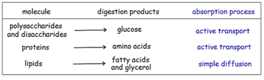
6.1.U6 Different methods of membrane transport are required to absorb different nutrients.
- List four methods of membrane transport required to absorb nutrients.
- Describe the absorption of triglycerides.
- Describe the absorption of glucose.
Secondary Active Transport
- A transport protein couples the active translocation of one molecule to the passive movement of another (co-transport)
- Glucose and amino acids are co-transported across the epithelial membrane by the active translocation of sodium ions (Na+)
Facilitated Diffusion
- Channel proteins help hydrophilic food molecules pass through the hydrophobic portion of the plasma membrane
- Channel proteins are often situated near specific membrane-bound enzymes (creates a localised concentration gradient)
- Certain monosaccharides (e.g. fructose), vitamins and some minerals are transported by facilitated diffusion
Osmosis
- Water molecules will diffuse across the membrane in response to the movement of ions and hydrophilic monomers (solutes)
- The absorption of water and dissolved ions occurs in both the small and large intestine
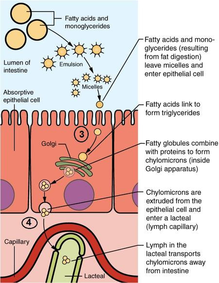
Simple Diffusion
- Hydrophobic materials (e.g. lipids) may freely pass through the hydrophobic portion of the plasma membrane
- Once absorbed, lipids will often pass first into the lacteals rather than being transported via the blood
During absorption nutrients from food must pass from the lumen of the small intestine to the cells in the capillaries or lacteals in the villi.
- Many types of transport are used to move different nutrients into and out of the epithelium cells of the villi
These modes of transport will be outlined using two products of digestion.
Glucose
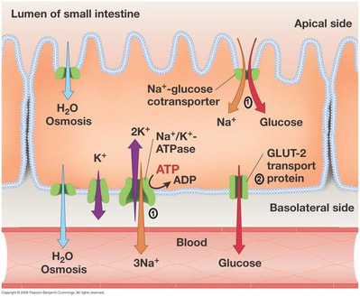
Since glucose has many hydroxyl groups it is a polar molecule and cannot pass through the cell membrane by simple diffusion and therefore relies on different types of facilitated diffusion in order to move into and out of the epithelial cells of the villi. (The numbers 1-4 here, match with numbers on the two diagrams above and below).
1) As seen above Na+ is pumped out of the cytoplasm of the epithelial cells into the interstitial space inside the interstitial by sodium/potassium pumps.
- This creates a concentration gradient between the lumen and the cytoplasm of the epithetical cells.
- This means Na+ ions want to diffuse into the epithelial cells.
- Co-transport proteins in the membrane of the microvilli, allow a sodium ion and a glucose molecule to be transported together into the epithelial cell.
This type of facilitated diffusion is passive, but requires active transport of the Na+ ions out of the cell to create the concentration gradient.
2) Specific glucose channels allow glucose to diffuse from the epithelial cells into the blood cells of the capillaries
3) Fatty acids, glycerol and monoglycerides, which are products of lipid digestion can diffuse into epithelial cells from the lumen by passive diffusion
4) Inside the epithelial cells, fatty acids and monoglycerides reform into triglycerides, and therefore can’t move back out into the lumen because of their size. These lipids combine together with proteins and phospholipids to form lipoproteins. The lipoproteins are then excreted by exocytosis, enter the lacteal and are carried away by the lymph
6.1.A1 Processes occurring in the small intestine that result in the digestion of starch and transport of the products of digestion to the liver. (Oxford Biology Course Companion page 285)
- Describe the structure of starch.
- Outline the source, function and specificity of amylase.
- Outline the digestion of maltose, maltotriose and dextrins into glucose.
- Describe absorption of glucose by villus epithelial cells.
- Describe transport of glucose into and through villi capillaries.
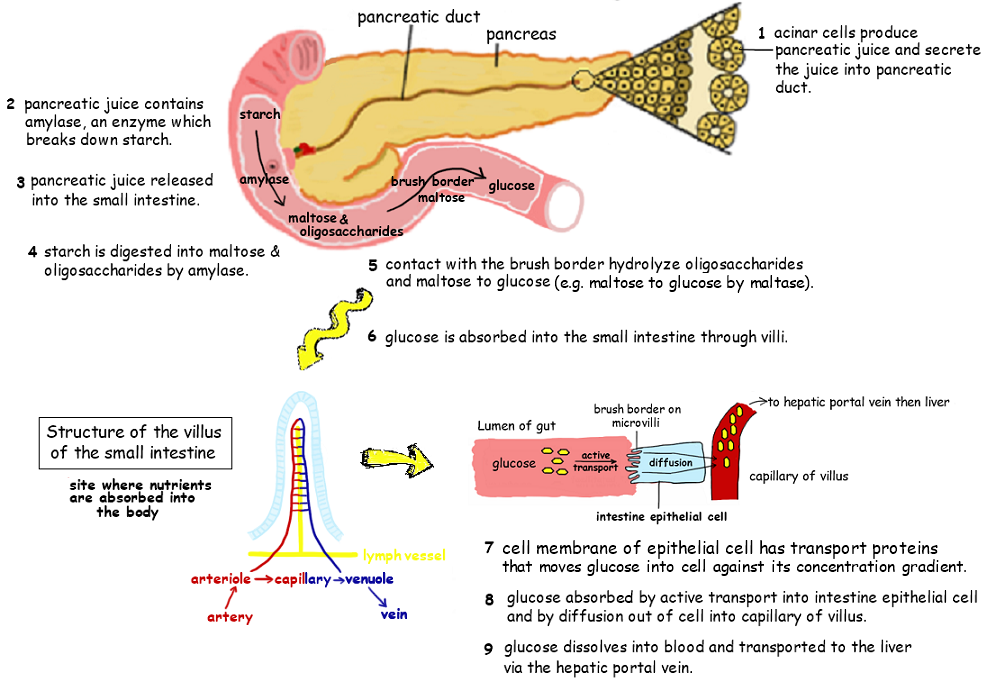
Starch is a polysaccharide composed of glucose monomers and accounts for ~ 60% of the carbohydrates consumed by humans. Starch can exist in one of two forms – linear chains (amylose) or branched chains (amylopectin)
- Many catabolic reactions take place in the small intestine
- These reactions are catalyzed by a number of enzymes that break down starch into smaller disaccharides and trisaccharides, which are further broken down into monosaccharides.
- These reactions need to occur since the starch molecule is much too large to pass through the membranes of the small intestine
- Starch-a long chain of α (alpha) glucose molecules used as a glucose storage by plants
- Starch consists of two types of molecules, amylose which linear and amylopectinwhich is branched (look at your notes from topic 2)
- Amylose 1,4 bonds can be broken apart by amylase to form the disaccharide maltose and the trisaccharide maltotriose
- Amylose cannot break the 1,6 bond seen in amylopectin
- These larger fragments containing this 1,6 bond of amylopectin are called dextrins
- These dextrins are further broken down by another enzyme into maltose
- Finally, all the maltose is hydrolyzed into glucose by maltase in order for it to be transported from the lumen of the small intestine into the blood in the capillaries surround the small intestine
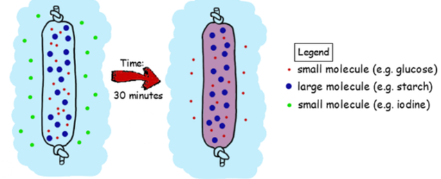
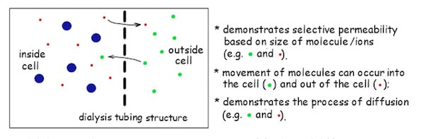
6.1.A2 Use of dialysis tubing to model absorption of digested food in the intestine.
- Explain the use of dialysis tubing as a model for the small intestine.
The process of digestion therefore performs two key functions:
- It breaks down insoluble molecules into smaller subunits which can be readily absorbed into body tissues
- It breaks down inert molecules into usable subunits which can be assimilated by cells and reassembled into new products
The size-specific permeability of cell membranes can be modelled using dialysis tubing (Visking tubing)
- Dialysis tubing contains pores typically ranging from 1 – 10 nm in diameter and is semi-permeable according to molecular size
- Large molecules such as starch cannot pass through the tubing, however smaller molecules (such as maltose) can cross
- Unlike the membranes of living cells, dialysis tubing is not selectively permeable based on charge (ions can freely cross)
How dialysis tubing demonstrates absorption
- models are ideas used to explain things observed in the world
- models share imprortant characteristics with the phenomenon being demonstrated
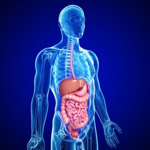
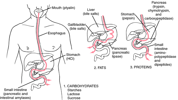
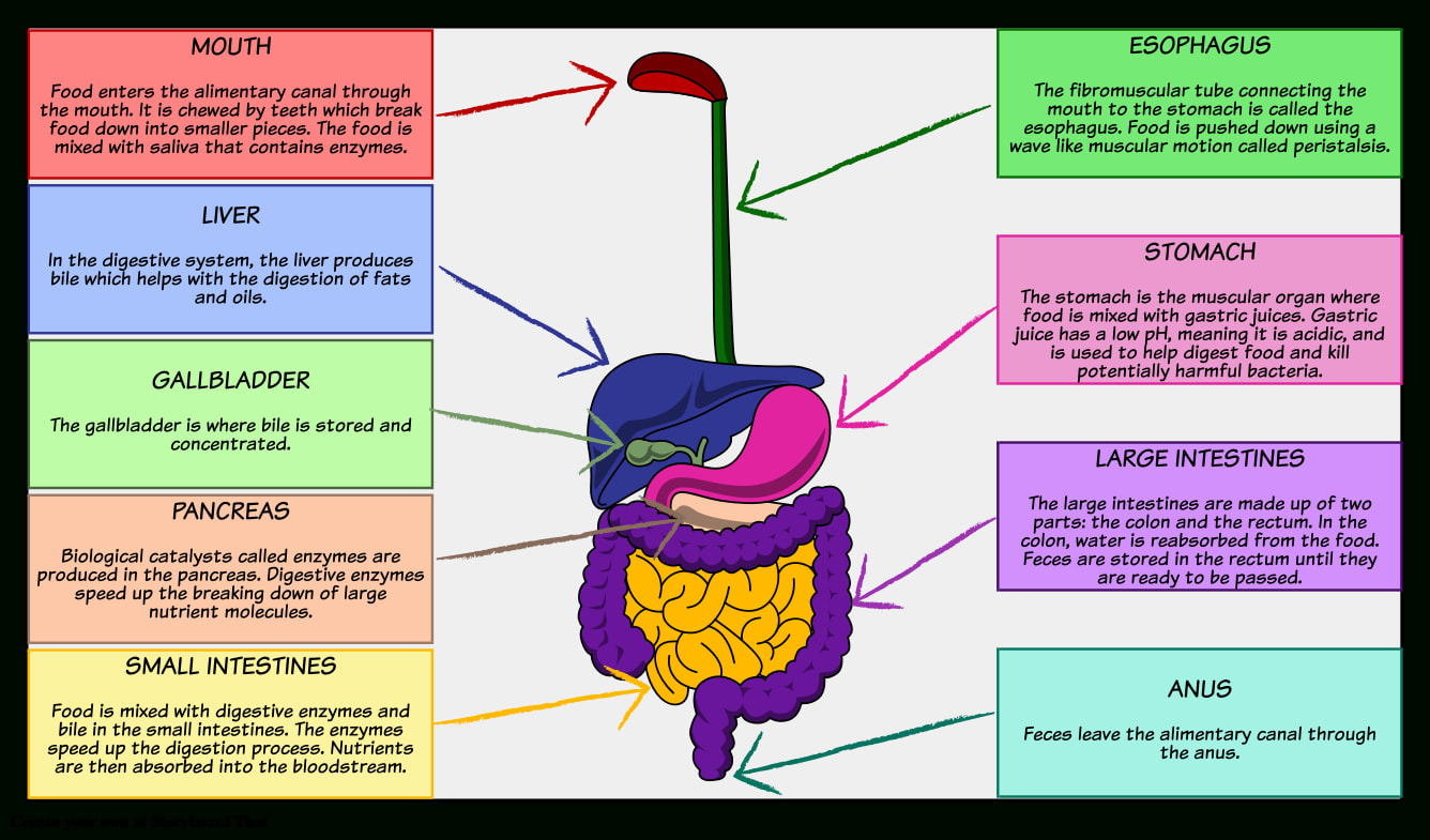
S 6.1.1 Production of an annotated diagram of the digestive system.
- State the role of the digestive system.
- Draw a diagram of the human digestive system.
- Outline the function of the following digestive system structures: mouth, esophagus, stomach, small intestine, pancreas, liver, gall bladder, and large intestine.
The human digestive system consists of an alimentary canal and associated accessory glands. The main parts to identify in the figure below include the mouth, esophagus, stomach, small intestine, large intestine and anus along with the digestive glands – liver and pancreas. Also include the gall bladder. Make note of the interconnections between these structures the functions of the stomach, small intestine and large intestine.
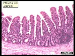
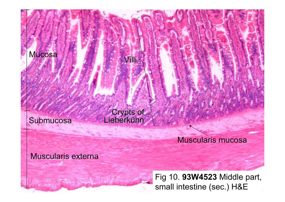
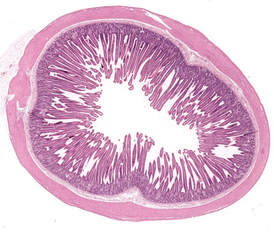
S 6.1.2 Identification of tissue layers in transverse sections of the small intestine viewed with a microscope or in a micrograph. (Oxford Biology Course Companion page 281
- Outline the function of the four layers of tissue found in the wall of the small intestine.
- Label the four layers of tissue found in the wall of the small intestine as viewed with a microscope or in a micrograph.
The small intestine is composed of four main tissue layers, which are (from outside to centre):
- Serosa – a protective outer covering composed of a layer of cells reinforced by fibrous connective tissue
- Muscle layer – outer layer of longitudinal muscle (peristalsis) and inner layer of circular muscle (segmentation)
- Submucosa – composed of connective tissue separating the muscle layer from the innermost mucosa
- Mucosa – a highly folded inner layer which absorbs material through its surface epithelium from the intestinal lumen
Longitudinal Cross Section of Small Intestine |
Transverse Cross Section of Small Intestine |
Digestion and Absorption
Table of Content
- What is Digestion?
- Types of Digestion
- Digestive system
- Digestion of food
- Digestion of Carbohydrates
- Digestion of fats
- Digestion of proteins
- Absorption of digested products
What is Digestion?
The process of breakdown of complex, water insoluble food molecules into simple water soluble molecules is known as Digestion. It is a complex process involving various enzymes, secretion etc.
Types of Digestion
Mechanical digestion is a process of physically breaking the food into smaller pieces. It includes the food that is chewed in the mouth.
Chemical digestion is complex process of digestion of food involving enzymes which breakdown complex food molecules into simple food molecules.
Digestive system
The human digestive system comprises of alimentary canal and digestive glands.
Alimentary canal
The anterior most part of the alimentary canal is known as mouth and the posterior most part is known as Anus. The mouth leads into the oral cavity or buccal cavity. Buccal cavity contains teeth and tongue. Each tooth is fixed in socket of jaw bond. This type of arrangement of teeth is known as Thecodont. For Example, Mammals.
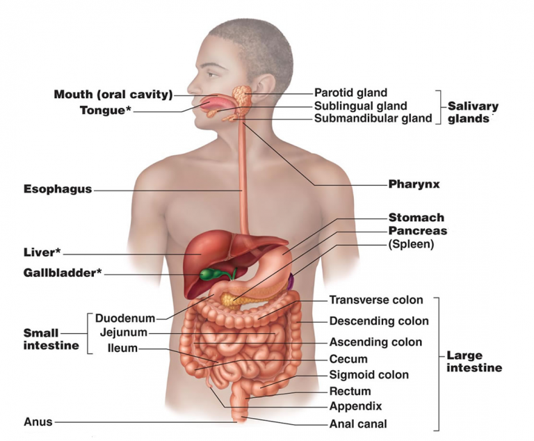
Fig. 1. Alimentary canal
In mammals, temporary teeth are replaced by permanent teeth in adults. This is known as Diphyodont. A human adult consists of 4 types of teeth which are 32 in number. The 4 types of teeth are- Incisors, Canines, Molars, and Premolars.
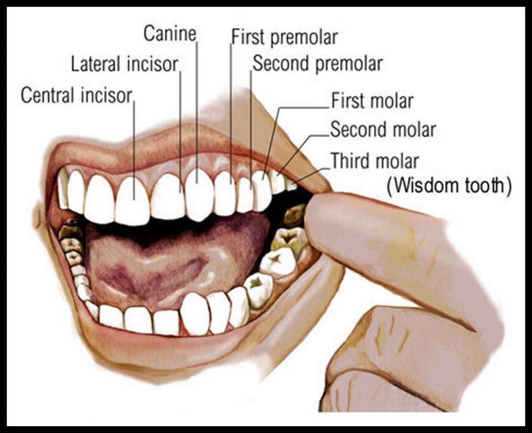
Fig. 2. Types of teeth
Surface of the teeth is covered by the hardest substance known as Enamel. The tongue is freely movable, muscular organ that helps in chewing of food inside the oral cavity. The oral cavity leads into the pharynx, which is common passage for food as well as air. Esophagus also known as Food Pipe helps in passage of the food to the stomach.
Stomach is a bag-like structure located in the upper portion of the abdominal cavity. It is divided into three parts – Cardiac, Fundic and Pyloric.
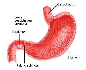
Fig. 3. Parts of stomach
Pyloric is the region which opens into the first part of the small intestine. Small intestine is divided into duodenum, jejunum and ileum. Pyloric sphincter guards the opening of stomach into the duodenum. Ileum opens into the large intestine.
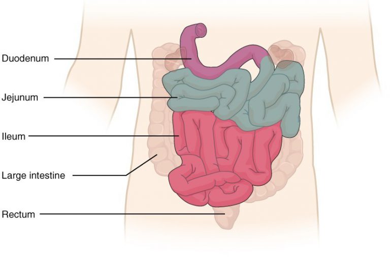
Fig. 4. Parts of small intestine
Large intestine is divided into caecum, colon, and rectum. Rectum finally opens into the anus. Symbiotic organisms reside in caecum. A vestigial organ known as Vermiform appendix arises from the caecum. Colon is divided into ascending, transverse, and descending part.
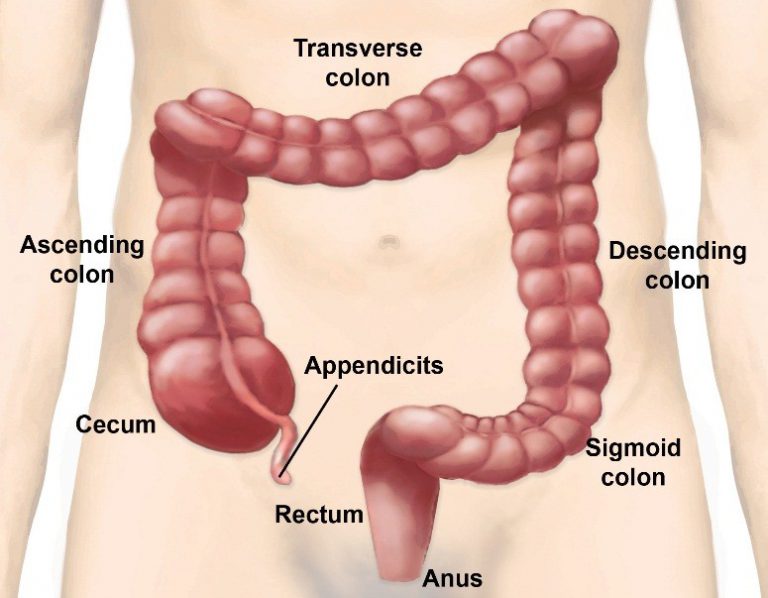
Fig. 5. Parts of large intestine
The wall of the alimentary canal starting from the esophagus to the rectum is lined by four layers- outermost layer serosa, inner to it lies the muscularis, next submucosa and innermost mucosa. Muscularis is made up of two types of muscles – Circular Muscles and Longitudinal Muscles. Mucosa for folds in the stomach and small finger-like projections in intestine known as Villi. Villi increases the surface area for efficient absorption of food.
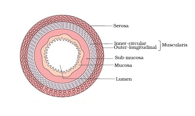
Fig. 6. Wall of the alimentary canal
Mucosa forms gastric glands in stomach, crypts in the intestine
Digestive glands
The three main digestive glands are – Salivary Glands, Liver and Pancreas.
There are three pairs of salivary glands present – the parotids glands located in cheek, the sub-maxillary/sub-mandibular located in lower jaw and the sublinguals, found below the tongue. These glands secrete salivary juice. Salivary juice contains an enzyme salivary amylase or ptyalin, that participates in digestion of carbohydrates which begin in the mouth.
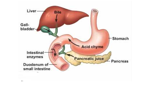
Fig. 7. Digestive glands
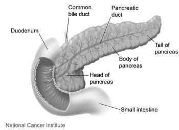
Liver is the largest gland found in the human body. The structural and the functional unit of liver is hepatic lobules. Each lobule is connected by a tissue known as Glisson’s Capsule. Liver secretes bile juice which is stored in muscular sac-like structure known as Gall Bladder. The duct of the gall bladder is known as Cystic Duct that combines with the duct of the liver to form common bile duct.
The bile duct and the duct of the pancreas open into duodenum.
Pancreas is an endocrine as well as exocrine gland. The endocrine function of the pancreas involves the secretion of insulin and glucagon which regulates the blood sugar level. Whereas endocrine function of pancreas includes secretion of pancreatic juice that participates in digestion of food.
Digestion of food
Digestion of food begins in the buccal cavity where two important functions are performed- mastication of food and swallowing. Teeth and tongue promote mastication of food. Saliva mixes the food thoroughly and forms bolus. The saliva in the mouth contains electrolytes such as K+, Na+ etc. and two important enzymes known as lysozyme and salivary amylase. Salivary amylase breakdown starch into maltose, a disaccharide.
The bolus is then passed to pharynx and then to esophagus. The movement of food inside the esophagus is known as Peristalsis. The gastro-esophageal sphincter guards the food into the stomach.
The mucosa in the stomach forms the gastric glands. Gastric glands consist of-
- Neck cells that secrete the mucus. Mucus lubricates the lining of the stomach against excoriation by hydrochloric acid.
- Peptic cells that secrete pepsinogen.
- Parietal or oxyntic cells that secrete hydrochloric acid, which creates acidic environment inside the stomach.
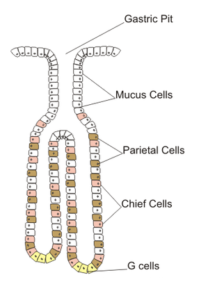
Fig. 8. Gastric gland
When food mixes with the stomach, it forms chyme. In presence of hydrochloric acid, pepsinogen is converted into pepsin. Pepsin is an enzyme that converts proteins into peptones and proteases.
Rennin is also secreted by gastric glands in infants which helps in digestion of milk proteins.
Small intestine receives secretions such as bile, pancreatic juice and intestinal juice. The pancreatic juice contains enzymes which are present in inactive form or pro-enzymes – Trypsinogen, Chymotrypsinogen, Procarboxypeptidases, Amylases, Lipases and Nucleases.
The intestinal mucosa has goblet cells that secretes mucus. The secretions of the brush border cells of the mucosa along with the secretions of the goblet cells forms the intestinal juice or succus entericus. It contains a variety of enzymes like disaccharidases (Example: Maltase), Dipeptidases, Lipases, Nucleosidases, etc.
Enterokinase, an enzyme secreted by intestinal mucosa activates trypsinogen to form trypsin.
Digestion of Carbohydrates
Digestion of Carbohydrates begins in the mouth. Salivary amylase helps in digestion of starch into maltose. Further digestion of carbohydrates occurs due to pancreatic amylase present in pancreatic juice in small intestine. Intestinal juice also contains enzymes such as maltase which digest maltose into glucose, sucrase which digest sucrose into glucose and fructose, lactase which digest lactose into glucose and galactose.
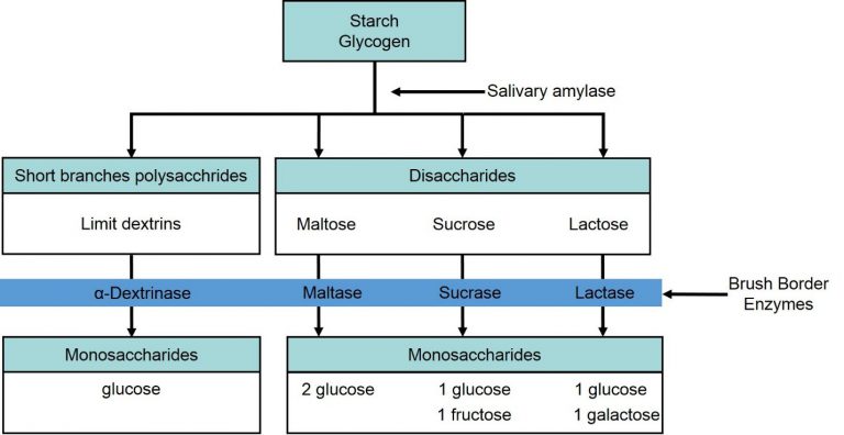
Fig. 9. Digestion of carbohydrates
Digestion of fats
Small amount of lipase is secreted by gastric glands where begins the process of fat digestion. Then the small intestine contains bile secreted by liver, which helps in emulsification of fats, that is, breakdown of fats into small micelles. Intestinal juice also contains lipases that aids in digestion.
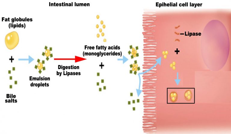
Fig. 10. Emulsification of fats
Digestion of proteins
This process begins in stomach, where, pepsin breaks proteins into peptones and proteases. Trypsin, chymotrypsin, carboxypeptidase breakdown peptones, proteases into dipeptides.
The undigested and unabsorbed substances are passed to large intestine, from where they can be removed from the body. No important digestion process occurs in large intestine.
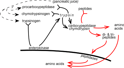
Fig. 11. Digestion of proteins
Absorption of digested products
The end products of digestion pass from intestine to the blood or lymph for absorption. Glucose, amino acids, electrolytes, are absorbed by simple diffusion. Some amino acids and glucose are transported with the help of carrier molecules, this type of transport is known as Facilitated Transport.
Transport of ions such as sodium ions occurs via active transport. Fatty acids and glycerol which are end products of fats are insoluble in water, so first gets converted into micelles which move into intestinal mucosa.
Maximum absorption occurs in small intestine.