Topic 7.3: Translation
In the Translation Unit we will extend our knowledge on Topic 2.7 DNA Replication, Transcription and Translation.
Translation is the second part of the central dogma of molecular biology, describing how the genetic code is used to make amino acid chains. This unit will explore the mechanics involved in polypeptide synthesis. You will learn the three major steps of translation as tRNA, mRNA, and ribosomes form the polypeptide. We will also explore the structural formation of polypeptides after translation
Essential idea:
- Information transferred from DNA to mRNA is translated into an amino acid sequence.
Nature of science:
- Developments in scientific research follow improvements in computing—the use of computers has enabled scientists to make advances in bioinformatics applications such as locating genes within genomes and identifying conserved sequences. (3.7)
7.3 U 1 Initiation of translation involves assembly of the components that carry out the process.
- Outline the process of translation initiation.
The first stage of translation involves the assembly of the three components that carry out the process (mRNA, tRNA, ribosome)
Start codon:
- the mRNA triplet codon AUG is universally the start codon used to mark the beginning of the coding sequence of a gene;
- the tRNA with the anticodon UAC, and carrying the amino acid methionine, is always the first tRNA to enter the P-site during translation
Initiation:
- 5’ end of mRNA binds to the small subunit of the ribosome
- initial mRNA codon = AUG = start codon
- tRNA with anticodon: UAC binds to mRNA AUG codon by complementary base pairing, methionine attached to tRNA 3’ terminal
- large ribosomal subunit binds, completing ribosomal structure, and producing two ribosomal binds sites: P site & A site

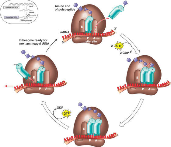
7.3 U 2 Synthesis of the polypeptide involves a repeated cycle of events.
- Outline the process of translation elongation, including codon recognition, bond formation and translocation.
- State the direction of movement of the ribosome along the mRNA molecule.
Elongation
- While the first tRNA is still attached, a second tRNA attaches to the mRNA at the A site on the ribosome, carrying the amino acid that corresponds to the mRNA codon.
- The methionine amino acid at the P site binds to the amino acid carried by the second tRNA located at the A site.
- The two amino acids are joined together through a condensation reaction that creates a peptide bond between the two amino acids.
Translocation
- The ribosome moves along the mRNA one codon shifting the tRNA that was attached to methionine to the E site.
- The tRNA is released back into the cytoplasm from the E site, allowing it to pick up another amino acid (methionine) to build another polypeptide.
- Another tRNA moves into the empty A site bringing the next amino acid corresponding to the mRNA codon.
- Again, the amino acid is attached to the polypeptide forming a peptide bond, the ribosome slides across one codon and tRNA at the P site moves into the E site releasing it back into the cytoplasm.
- The ribosome continues to move along the mRNA adding amino acids to the polypeptide chain.
- This process continues until a stop codon is reached.
7.3 U 3 Disassembly of the components follows termination of translation.
- Outline the process of translation termination, including the role of the stop codon.
Termination:
- when ribosomal A-site reaches a stop codon, no tRNA has a complementary anticodon
- there are three stop codons in the genetic code; none of these have a corresponding tRNA
- when a ribosome encounters a stop codon, a release factor binds to the stop codon
- release factor protein binds to ribosomal A-site stop codon
- polypeptide and mRNA are released
- large and small ribosomal subunits separate
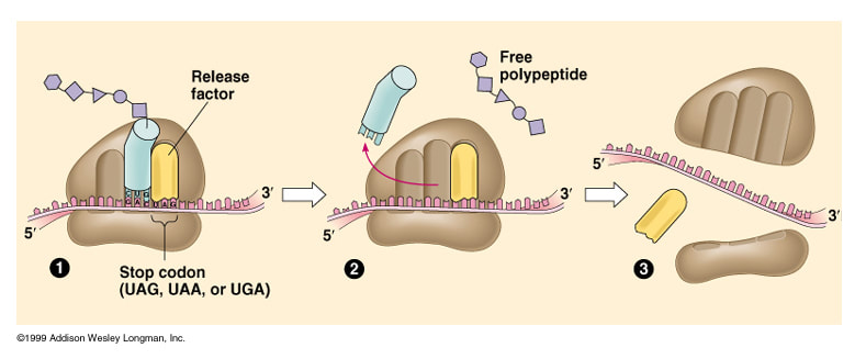
- Ribosome reaches the STOP codon. Release factor attaches
- tRNA released to find another amino acid. Polypeptide released
- Components of the ribosomoe break apart. All are used again
7.3 U 4 Free ribosomes synthesize proteins for use primarily within the cell.
- State the difference between free and bound ribosomes.
- List destinations of proteins synthesized on free ribosomes.
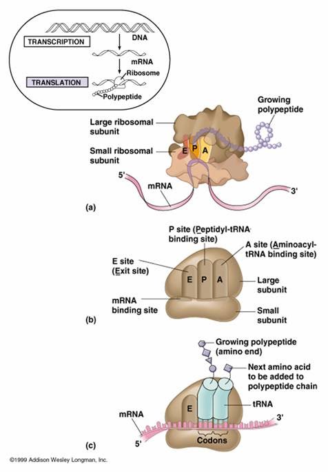
Ribosomes are composed of rRNA and protein.
- protein (40% of the weight) and rRNA (60% of weight) composition:
- large and small subunits
- three tRNA binding sites: E-site, P-site, A-site
- mRNA binding site: on small ribosomal subunit
Free ribosomes are located in the cytoplasm, and the bound ribosomes are attached to the endoplasmic reticulum. Free ribosomes produce proteins for the cell, while bound ribosomes produce proteins that are transported out of the cell.
Free ribosomes in the cytoplasm synthesize proteins that will be used inside the cell in the cytoplasm, mitochondria and chloroplasts (in autotrophs).
Free ribosomes synthesize proteins for use primarily within the cell itself that bound ribosomes synthesize proteins primarily for secretion or for lysosomes
7.3 U 5 Bound ribosomes synthesize proteins primarily for secretion or for use in lysosomes. (Oxford Biology Course Companion page 366).
- List destinations of proteins synthesized on bound ribosomes.
- Outline how a ribosome becomes bound to the endoplasmic reticulum.
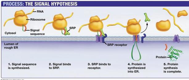
Proteins perform many functions within specific compartments of the cell or in other parts of the body after they are secreted out of the cell
Proteins that are destined to be used in lysosomes, ER, Golgi Apparatus, the plasma membrane or secreted by the cell are made by ribosomes bound by the endoplasmic reticulum. Ribosomes that become bound to the ER are directed here by a signal sequence that is part of that specific polypeptide. This signal sequence on the polypeptide binds to a signal recognition protein (SRP). The SRP guides the polypeptide and ribosome to the ER where it binds to an SRP receptor. Translation can now continue and the polypeptide is deposited into the lumen of the ER as its created for transportation to the correct location
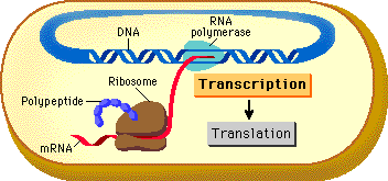
7.3 U 6 Translation can occur immediately after transcription in prokaryotes due to the absence of a nuclear membrane.
- Compare the timing and location of transcription and translation between prokaryotes and eukaryotes.
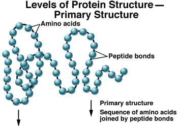
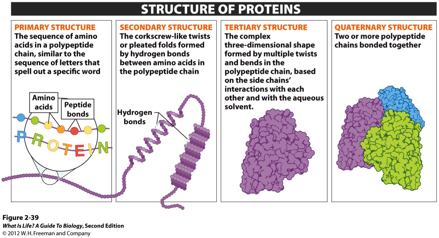
7.3 U 7 The sequence and number of amino acids in the polypeptide is the primary structure.
- Describe the primary structure of a protein, including the type of bonding involved.
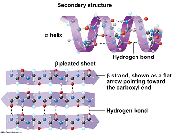
7.3 U 8 The secondary structure is the formation of alpha helices and beta pleated sheets stabilized by hydrogen bonding.
- Describe the secondary structure of a protein, including the type of bonding involved.
- Identify the alpha-helix and beta-pleated sheet in images of protein structure.
Weak hydrogen bonds between amino and carboxyl groups and different amino acids form at regular intervals, creating a regular structure (not from interactions between variable R- groups)
- alpha helix = coiling into a helix
- beta pleating = a folded sheet as polypeptide folds back on itself
- not all of a polypeptide forms secondary structure in most proteins
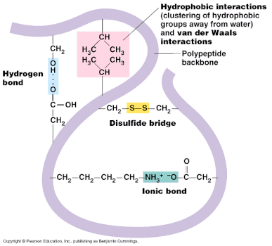
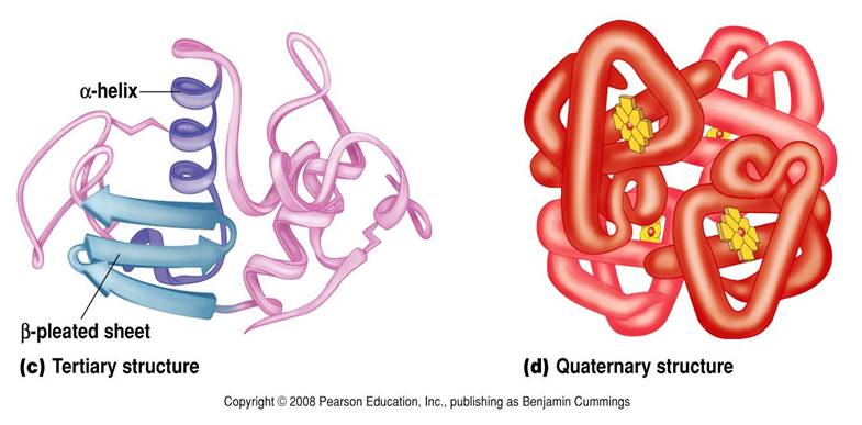
7.3 U 9 The tertiary structure is the further folding of the polypeptide stabilized by interactions between R groups.
- Describe the tertiary structure of a protein, including the types of R group interactions involved.
- Explain how the chemical characteristics of R groups in the polypeptide chain affect protein folding.
Tertiary structure is the third level of protein organization. Tertiary structure develops a three dimensional shape because of the interactions that occur between the R groups.
- Disulphide bridges form between sulphur atoms on the R-groups from the amino acid cysteine.
- Hydrogen bonds form between polar R-groups and other polar R-groups.
- Ionic Bonds are also formed between some R-groups
- Hydrophobic R-groups turn inwards away from water and hydrophilic R-groups face outwards towards the water.
- Protein is globular in nature.
- Interactions between variable R- groups forming:
- hydrophobic interactions between nonpolar amino acids
- hydrogen bonds between polar amino acids
- ionic bonds between ionic amino acids
- covalent bonds between sulfur containing amino acids
- Producing the three-dimensional folded structure of most proteins
7.3 U 10 The quaternary structure exists in proteins with more than one polypeptide chain.
- Outline the quaternary structure of protein folding.
- Describe the structure of a conjugated protein, including the prosthetic group.
Protein consisting of more than one polypeptide chain. These chains can consist of quaternary structures. Quaternary structure exists in many proteins such as hemoglobin (4 polypeptide chains).
- Hemoglobin also contains a prosthetic group (non-polypeptide structure). The prosthetic group in hemoglobin is iron. Hemoglobin contains 4 iron molecules.
Proteins with prosthetic groups are called conjugated proteins. Aggregations of polypeptides form interactions between more than one polypeptide
- polypeptides + non-proteinaceous molecules, such as:
- metals, e.g. iron in hemoglobin
- vitamins as enzyme cofactors
- nucleic acids as in ribosomes
- carbohydrates in glycoproteins
- lipids in lipoproteins
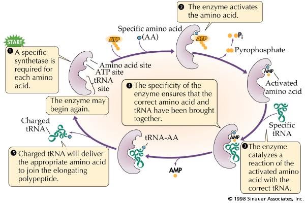
7.3 A 1 tRNA-activating enzymes illustrate enzyme–substrate specificity and the role of phosphorylation.
- State the role of the tRNA activating enzymes.
- Outline the process of attaching an amino acid to tRNA by the tRNA activating enzyme.
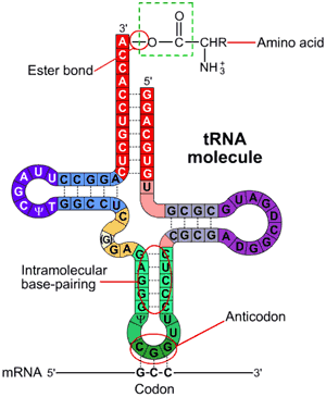
There are many different types of tRNA in a cell.All have tRNA molecules have:
- a triplet of bases called the anticodon, found in the anticodon loop of 7 nucleotides
- two other loops
- a CCA base sequence at the 3’ terminal, which forms a site for attaching an amino acid d) sections that become double-stranded by base pairing
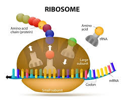
- Describe the structure of the ribosomes, including the small and large subunits and the names and roles of the tRNA binding sites.
- Use molecular visualization software to view and identify the small and large subunit and tRNA binding sites of the ribosome.
- Outline the structure of tRNA molecules.
- Use molecular visualization software to view and identify the anticodon and amino acid binding site of a tRNA.
Ribosomes are organelles made from protein and RNA that catalyze the assembly of amino acids into polypeptides during translation. Ribosomes consist of two subunits; a large subunit and a small sub unit. The large sub-unit consists of 3 binding sites for tRNA molecules called the A site (The A site binds an aminoacyl-tRNA (tRNA bound to an amino acid); the P site binds a peptidyl-tRNA (tRNA bound to the peptide being synthesized); and the E site binds a free tRNA (no amino acid attached) before it exits the ribosome. The order of the sites in the ribosomes is E P A.
There is a binding site for the mRNA on the surface of the ribosome The small subunit of the ribosome contains the binding site for the mRNA strand. The space between the two subunits of the ribosome is where the polypeptide is assembled during translation.
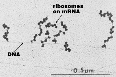
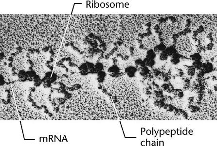
7.3 S 2 Identification of polysomes in electron micrographs of prokaryotes and eukaryotes. (Oxford Biology Course Companion page 366).
- Outline the structure of a polysome.
- Identify the beginning of an mRNA strand in a micrograph of polysomes.
In prokaryotes, several ribosomes can attach themselves to the growing mRNA chains to form a polysome while the mRNA chains are still attached to the DNA (left)
In eukaryotes, the mRNA detaches from the DNA and is then transported through pores in the nuclear envelope to the ribosomes in the cytoplasm. Once in the cytosol, eukaryote mRNA can also form polysomes (right)